


FIG. 10.
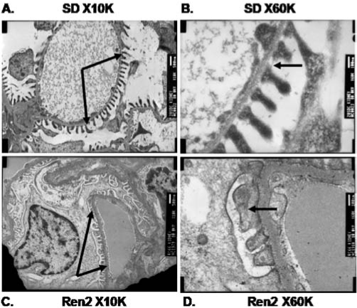
Ultrastructural structural depiction of podocyte foot processes in the transgenic TG(mRen2)27 Ren2 vs. Sprague-Dawley (SD) rats with transmission electron microscopy. In the SD rats, the podocytes form uniform processes along the basement membrane (arrows, top left A) with a prominent slit-pore diaphragm present (arrow, top right B). In the Ren2 rat (arrows, bottom left C), the podocytes have lost their nice uniformity with effacement (arrow, bottom right D).