


Fig. 7. Hypersensitivity of PARP-1−/− cells to camptothecin and functional Tdp1 deficiency in PARP-1 −/− cells.
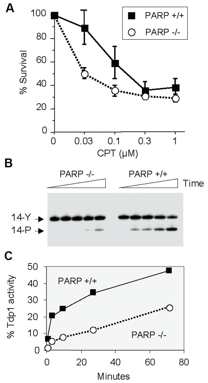
A. Mouse fibroblasts were exposed to camptothecin (concentrations indicated on X axis) for 1 hour, washed, kept in culture for 4 days and counted. The curves represent the averages and standard deviations from five independent experiments. B. A 14-mer single-stranded oligonucleotide with a phosphotyrosine at the 3′-end (see Fig. 6) (14-Y) [106] was incubated with nuclear extracts obtained from PARP-1−/− and PARP-1 +/+ cells [195] in the presence of 50 mM EDTA and absence of MgCl2 to eliminate PNKP activity, which requires MgCl2. A representative experiment is shown. TDP1 activity was determined as a shift in band position (from 14-Y to 14-P). C. Quantitation of the results shown in panel B using ImageQuant (Molecular Dynamics, Sunnyvale, CA).