


Figure 5. Examples of disorder-to-order transitions in β- and ι-MoRFs.
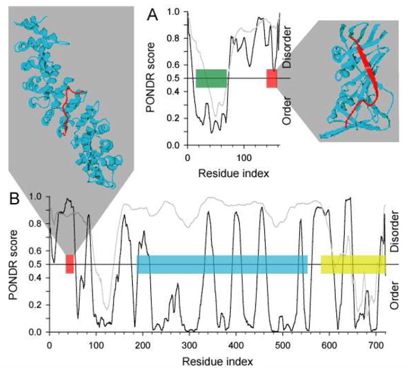
The structures of MoRFs (red ribbons) bound to their respective partners (blue ribbons) are shown. PONDR VL-XT predictions for the entire proteins in which these MoRFs are found are also shown, where the position of the MoRFs in these sequences are indicated (red boxes). (A) A β-MoRF example from p21 bound to PCNA (PDB entry 1AXC), where the position of the CDK inhibitor domain is indicated (green box). (B) An ι-MoRF example from Nup2p bound to karyopherin Kap60 (PDB entry 1UN0), where the positions of the FXF(G) repeat region (blue box) and the Ran binding domain (yellow box) are indicated.