


Fig. 3.
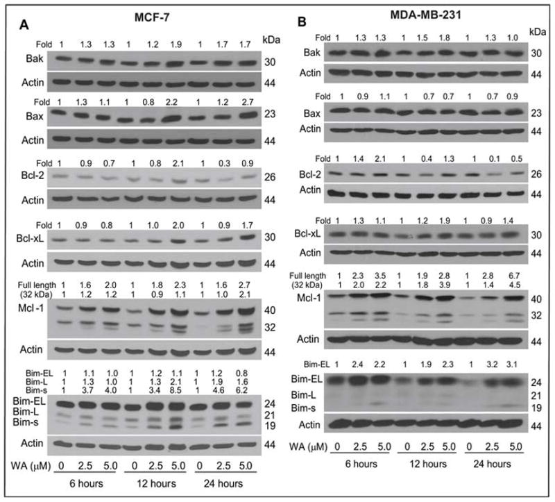
Immunoblotting for Bcl-2 family proteins using lysates from (A) MCF-7 cells and (B) MDA-MB-231 cells treated for 6, 12 or 24 h with DMSO (control) or the indicated concentrations of WA. The blots were stripped and re-probed with anti-actin antibody to correct for differences in protein loading. Changes in protein levels after correction for actin loading control are shown on top of the immunoreactive bands.