


Figure 7.
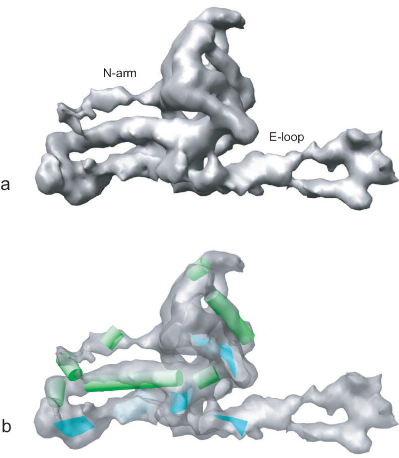
The secondary structure elements in the segmented subunit of ε 15 bacteriophage 3D density map. (a) The segmented subunit, in which labeled N-arm and E-loop region densities were well resolved at this resolution. (b) Seven α helices (in green) and two β sheets (in cyan) were identified by SSEHunter in the segmented subunit. The subunit density in (b) is displayed with some transparency.