


Figure 4.
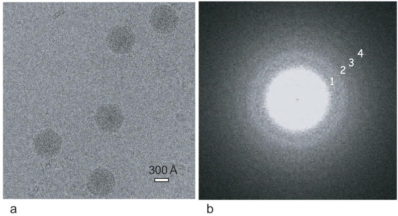
(a) One selected area (1560 pixel × 1560 pixel) from a typical cryo-image of ε 15 bacteriophage recorded on the CCD at an effective magnification of 56,000 × in a 300-kV electron cryomicroscope. The defocus for the image is ~1.55 μm. (b) The Fourier transform of 14 boxed-out particles from the whole CCD frame, portion of which is shown in (a). Four CTF zeros are marked with 1, 2, 3, 4, representing 17.8, 12.5, 10.1 and 8.8 Å, respectively.