


Figure 5. Constitutive Notch signaling antagonizes angiogenic sprouting mediated by MAGP-2 in MB114 cells.
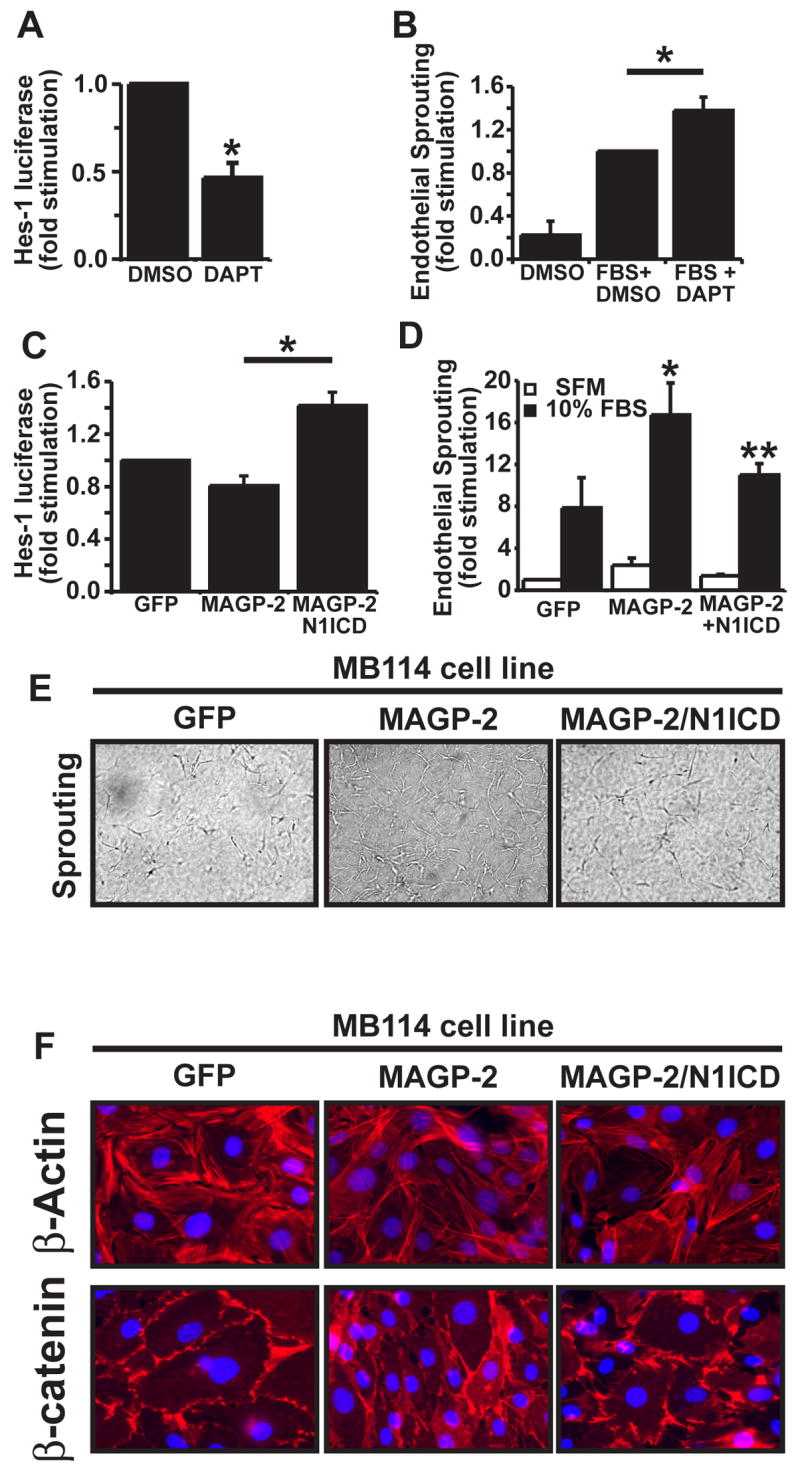
(A) MB114 cells were transiently transfected with pHes1-luciferase and pCMV-β-gal cDNAs, and subsequently were incubated overnight in the absence or presence of DAPT (10 μM). Afterward, luciferase and β-gal activities were determined. Data are the mean (± SEM) of two independent experiments. *, p<.05; student’s T-Test). (B) Quiescent MB114 cell monolayers were overlaid with rat tail collagen matrices, and were induced to form angiogenic sprouts by addition of 10% FBS supplemented with or without DAPT (10 μM). Five days later the number of invading angiogenic spouts were quantified by manual counting on a light microscope. Data are the mean (± SEM) of four independent experiment. (*, p < 0.05; Student’s T-Test). (C) GFP-, MAGP-2-, and MAGP-2/N1ICD-expressing MB114 cells were transiently transfected with pHes1-luciferase and pCMV-β-gal cDNAs. Luciferase and β-gal activities were determined 48 h post-transfection. Data are the mean (± SEM) of two independent experiments. (D) Quiescent monolayers of GFP-, MAGP-2-, and MAGP-2/N1ICD-expressing MB114 cells were overlaid with rat tail collagen matrices containing 10% FBS for 5 days. Afterward, the number of invading angiogenic sprouts were determined by manual counting under a light microscope. Data are the mean (± SEM) of four independent experiments. (*, **, p < 0.05; Student’s T-Test). (E) Quiescent monolayers of GFP-, MAGP-2-, and MAGP-2/N1ICD-expressing MB114 cells were either left untreated (quiescent) or overlayed with rat tail collagen matricies containing 10% FBS (sprouting) to induce angiogenic cell sprouting. After 5 days, angiogenic sprouts (100X magnification) were counted under a light microscope. (F) Quiescent monolayers of GFP-, MAGP-2-, and MAGP-2/N1ICD-expressing MB114 cells were prepared for immunofluorescent microscopy by incubating with rhodamine-conjugated phalloidin (upper panel) to visualize actin fibers (red) and DAPI to visualize nuclei (blue), or with β-catenin monoclonal antibodies (lower panel) to visualize cell-cell junctions (red) and DAPI (blue). Representative images are shown at 630X magnification from a single experiment that was conducted three times in its entirety.