


Figure 1. TLR4 deficient mice show decreased neuronal damage in the HF after GCI/R.
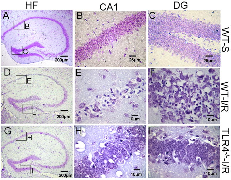
Brain sections were stained with 0.1% cresyl violet. Viable neurons were defined as neurons in which a normal nucleus can be seen. Damaged neurons exhibit features including pyknosis, karyorhexis and shrunken cell bodies. Three days after GCI/R, neuronal damage was prominent in the CA1 and dentate gyrus (DG) of the HF in WT mice. Compared with WT mice, there was less neuronal damage in the HF of TLR4-/- mice. A, D and G: HF; B, E and H: CA1; C, F and I: dentate gyrus (DG). WT-S: wild type sham, WT-I/R: wild type subjected to GCI/R, TLR4-/- -I/R: TLR4-/- mice subjected to GCI/R.