


Figure 2. EYFP-Stim1 colocalizes with α-tubulin.
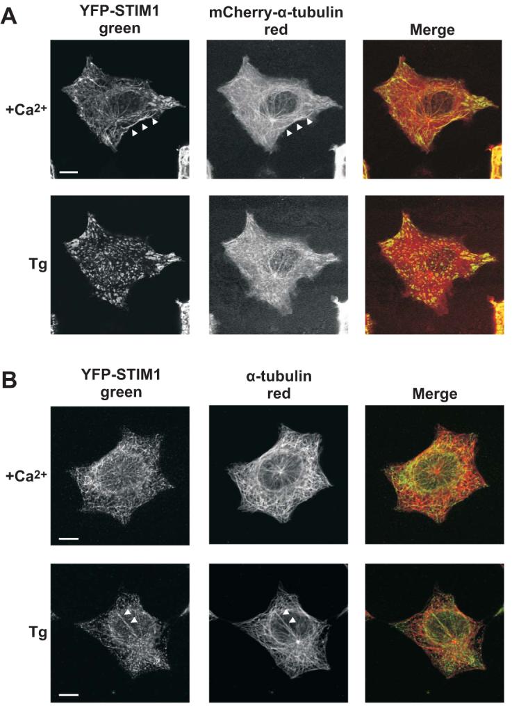
A) Upper panel: confocal images of an HEK293 cell co-overexpressing EYFP-Stim1 (left panel; green) and mCherry-α-tubulin (middle panel; red) in the presence of 1.8 mM extracellular Ca2+. The arrowheads indicate a track of EYFP-Stim1 fluorescence that exactly matches a corresponding track of mCherry-α-tubulin fluorescence. Lower panel: EYFP-Stim1 and mCherry-α-tubulin localization 15 min following store depletion with thapsigargin (Tg; 2 μM) in nominally Ca2+-free extracellular solution. B) Upper panel: Confocal images of a EYFP-Stim1 overexpressing HEK293 cell that was fixed and immunostained with antibodies against EYFP (to enhance detection of EYFP-Stim1) (left panel; green) and α-tubulin (middle panel; red). Lower panel: An HEK293 cell that was store depleted with thapsigargin for 15 min prior to fixation and immunostaining. Arrowheads indicate a track of α-tubulin along which several EYFP-Stim1 punctae appear to associate. Scalebars = 10 μm.