


Figure 3. Knocking Down EGFR Leads to Decreases in Intracellular Glucose Level, and Increasing the Glucose Content in the Culture Medium Rescues the Cell from Death Caused by Knocking Down EGFR.
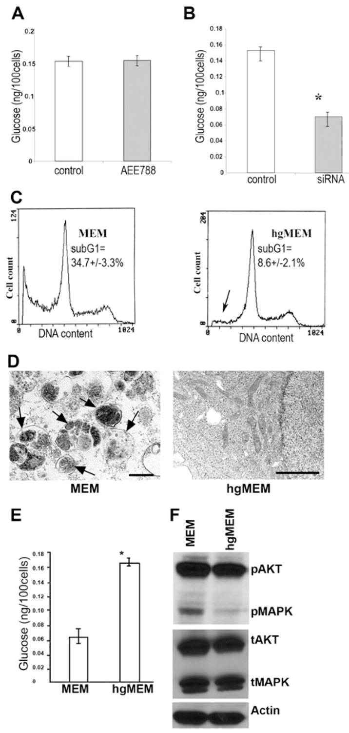
(A) Measurement of intracellular glucose content. AEE788 (5.0 μM) treatment for 72 hr did not alter the intracellular glucose content. (Error bars indicate mean ± SD.)
(B) The glucose content of EGFR siRNA-treated cells was significantly decreased (p < 0.001) compared with that of control cells after a 72 hr culture in MEM with 5.5 mM glucose. (Triplicate samples were used in each group. The asterisk indicates a significant difference with p < 0.001. Error bars indicate mean ± SD.)
(C) Culturing EGFR siRNA-transfected cells in high glucose (25 mM) MEM (hgMEM) significantly reduced (p < 0.01) the cell death proportion, as indicated by the decrease in sub-G1 cells (arrow). (Triplicate samples were used in this experiment.)
(D)The hgMEM-treated cells also lead to disappearance of autophagosomes on transmission electron microscopy (bar, 2 μm).
(E) The intracellular glucose level was also restored by hgMEM treatment. (Error bars indicate mean ± SD.)
(F) Western blotting shows that hgMEM treatment was also able to inhibit the enhanced activation of AKT and MAPK induced by knocking down EGFR.