


Figure 3.
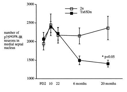
Analysis of the number and size of p75NGFR-IR neurons in the medial septal nucleus of Ts65Dn and 2n mice during development and in the adult. The mean cross-sectional profile area of p75NGFR-IR neurons with a nucleolus were assessed in Ts65Dn and 2n mice. No significant differences were found at PD2, PD10, or PD22. At later times, areas were as follows: (mean ± SEM) 6 months, Ts65Dn, 149 ± 2.2 (n = 5, n = 300) and 2n, 156 ± 2.3 (n = 5, n = 300), P = 0.04; 20 months, Ts65Dn, 147 ± 0.5 (n = 5, n = 506) and 2n, 153 ± 0.3 (n = 5, n = 506), P < 0.001.