


Figure 4. Pax6 is sufficient to promote Reelin expression and repress Slit1 expression in astrocytes.
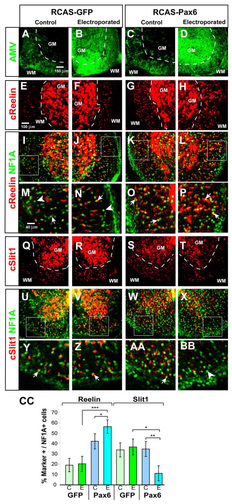
(A–D) Immunostaining for RCAS retroviral coat protein AMV in E12 spinal cord of chick embryos electroporated with either RCAS-GFP (A, B) or RCAS-Pax6 (C, D “WM,” white matter; “GM,” gray matter. (E-BB) Antibody staining for NFIA combined with fluorescent in situ hybridization for Reelin (E–P) or Slit1 (Q-BB) mRNAs. (I–L) and (U–X) are the same panels as in (E–H) and (Q–T), respectively, but include NF1A expression. (M–P) and (Y-BB) are higher power images of the boxed areas indicated in (I–L) and (U–X), respectively. Arrows indicate double-positive cells, arrowheads NFIA single-positive cells. (CC) The percentage of Reelin+ NFIA+ cells is significantly different between the electroporated (E) sides of Pax6 and GFP embryos (***, p <.001), as well as between the E and contralateral (C) sides of Pax6 embryos (*, p=.02). The reduction in Slit1 expression on the E side of Pax6 embryos is significant with respect to both the C side (**, p = .002), and the E side of GFP controls (*, p = .009). mean±S.E.M., n= 5 embryos per condition, two independent experiments.