


FIGURE 3.
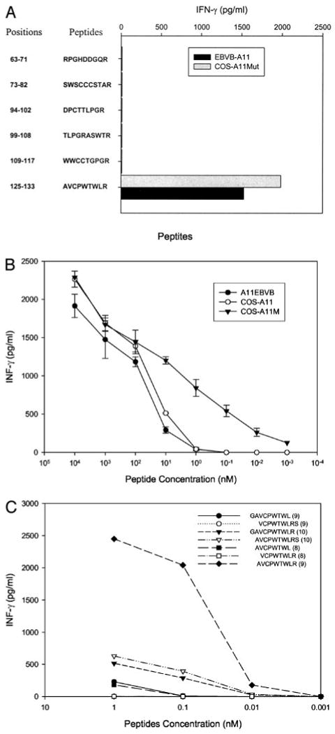
Identification and titration of the p16INK4a/p14ARF peptide recognized by clone T1D1. A, Peptide screening. Six peptides were synthesized and incubated for 2 h with A11-EBVB and COS-A11Mut cells at a concentration of 100 nM. Target cells were then cocultured with clone T1D1, and IFN-γ release was measured 18 h later. B, Peptide titration. Either HLA-A11-expressing EBVB, COS-A11N, or COS-A11Mut cells were loaded with the indicated concentrations of the AVCPWTWLR peptide for 2 h; clone T1D1 cells were added; and IFN-γ release was measured 18 h later. C, Testing of peptide variants. Variants of the peptide AVCPWTWLR either lacking or containing an additional N- or C-terminal residue were incubated with COS-A11Mut cells at indicated concentrations for 2 h. Clone T1D1 T cells were then added, and IFN-γ release was measured 18 h later.