


Figure 3. Effects of PCr on spontaneous Ca2+ waves in permeabilized myocytes.
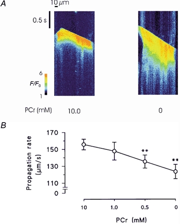
A, line scan images of Fluo-3 florescence during spontaneous Ca2+ waves in the presence and absence of PCr. Following withdrawal of PCr, spontaneous waves were markedly prolonged and the propagation rate was reduced. Both responses were from the same permeabilized myocyte. B, accumulated data showing changes in the propagation velocity of spontaneous Ca2+ waves following equilibration with solutions containing a range of [PCr]. Error bars represent the means ± s.e.m., n = 8. **P < 0.02.