


Figure 6. Timing of the input to the proximal region.
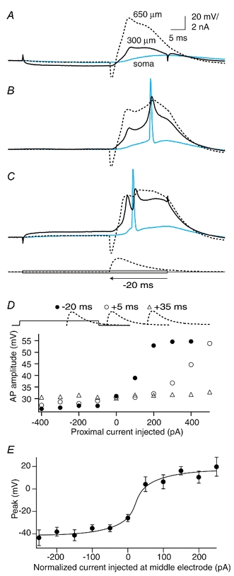
A, current injected at the proximal electrode (300 μm) caused the forward-propagating dendritic Na+-Ca2+ AP evoked by an EPSP-shaped waveform at the distal electrode (650 μm) to fail completely towards the soma. B, in this same cell the forward-propagating Na+-Ca2+ AP was in an intermediate state of propagation under control conditions and showed two components. The second rising component was interrupted by the presence of a back-propagating Na+ AP from the soma. C, depolarizing current at the proximal electrode caused the forward propagation to be completely active and only one component was then apparent. Further current injection led to more Na+ APs. D, the onset of an EPSP-shaped waveform injected at the distal electrode was moved relative to the end of the 50 ms step pulse injection at the proximal dendritic electrode. Here, we show data from 20 ms before the end of the proximal injection (•), and 5 ms (○) and 35 ms (▵) after the end of the proximal injection (shown above the graph schematically). The least current required to improve propagation of the dendritic Na+-Ca2+ AP was needed when it was injected during the proximal current injection (•). E, the sharp cutoff between APs that propagated well and those that propagated badly for a number of cells (n = 7) is shown by aligning the last point still under the half-amplitude point to 0 pA. The actual current injected for this point varied from -500 to +400 pA (mean, 100 ± 290 pA) depending on the initial conditions of coupling for the cell. The shift between good propagation and failure to propagate occurred within ≈50 pA.