


Figure 1.
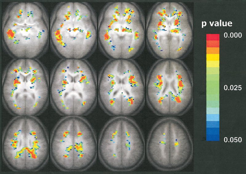
Composite map of the trace of the apparent diffusion coefficient negatively correlated with respect to the age of the subject. Colored pixels have P values less than .05 according to the Spearman rank correlation test, with a cluster size of 15, and are overlaid on the average (n = 33) T1-weighted transverse whole-brain data set. The colors indicate the level of statistical significance from a P value less than .05 (blue) to a P value less than .003 (red). Statistically significant decreases in the trace of the apparent diffusion coefficient with age are shown throughout the white matter. Single-shot spin-echo echo-planar diffusion-tensor MR imaging parameters were as follows: 3,250/80; matrix, 64 × 128; field of view, 19.2 × 25.6 cm; section thickness, 5 mm; Δ, 40 msec; δ, 15 msec. Modified driven-equilibrium Fourier transform whole-brain anatomic imaging parameters were as follows: 18/4.3; matrix, 256 × 128 × 96; field of view, 19.2 × 25.6 × 14.4 cm; τ, 550 msec.