


Figure 5.
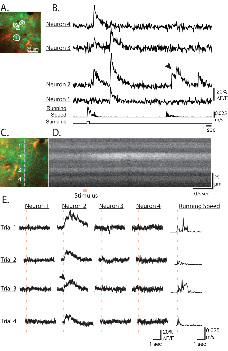
Line-scan and fast transient recordings in sensory cortex of awake mice. A) Image of neurons loaded with Calcium Green-AM (green channel) and astrocytes with SR101 (red channel). B) Fluorescence traces of the neurons shown in A from a time series recorded at 64ms/frame. C) Image of neurons loaded with Calcium Green-AM (green channel) and astrocytes with SR101 (red channel). The gray line represents the line-scanning position. D) Intensity versus time line-scan image (Trial 1) of the Calcium Green channel from the position shown in C during which an air puff stimulus was applied to the mouse. E) 4 trials of line-scan recorded fluorescence traces for the 4 neurons labeled in C. Traces are aligned to the stimulus timing (red dashed line). Mouse running speed is shown in the last column. Motion correction was not applied to the line scan recordings. Note the fast increases at the onset and the slower exponential offset decay of the fluorescence transients in all traces as well as the summation of transients in B) neuron 2 and E) neuron 2, trial 3 (arrowheads).