


Fig. 4.
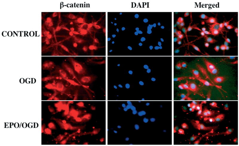
Eythropoietin (EPO) employs the Wnt pathway to modulate β-catenin. The pretreatment of EPO at 1 hour before oxygen-glucose deprivation (OGD) regulates the β-catenin location in microglia following OGD. Immunohistochemical double staining for β-catenin was obtained in microglia twelve hours following an six hour period of OGD by using a primary rabbit anti-β-catenin antibody with Texas-red avidin. OGD in microglia was performed by replacing media with glucose-free HBSS containing 116 mM NaCl, 5.4 mM KCl, 0.8 mM MgSO4, 1 mM NaH2PO4, 0.9 mM CaCl2, and 10 mg/L phenol red (pH 7.4) and cultures were maintained in an anoxic environment (95% N2 and 5% CO2) at 37 C for 6 hours. Cell nuclei were stained with DAPI (4′-6-diamidino-2-phenylinodole). Representative images illustrate the accumulation of β-catenin in the microglial cytoplasm during OGD and the translocation of β-catenin from the cytoplasm to nuclei following the application of EPO, but the expression of β-catenin exists in both cytoplasm and nuclei without cellular translocation in control (untreated) microglia.