


Figure 6.

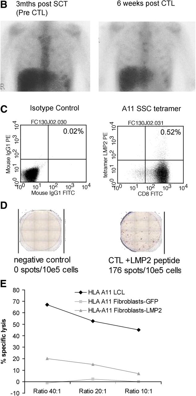
Tumor responses. (A) In patient 14 whose cervical lymph nodes responded to infusion of CTLs that were not genetically marked, immunohistochemical analysis revealed CD8+ tumor-infiltrating lymphocytes only after CTL infusion. This corresponded to the disappearance of LMP1+ cells. (B) A gallium scan demonstrating increased hilar uptake is observed in patient 12 3 mo after autologous SCT. The follow-up scan 6 wk after CTL infusion is reported as normal. (C) To demonstrate LMP2 specificity within patient 12's infused CTL line in vitro, the frequency of CTLs specific for the HLA-A11–restricted LMP2 epitope SSC was assessed using tetramer and (D) ELISPOT analysis, the latter using SSC peptide–pulsed PHA blasts as targets. (E) Further, the percent-specific 51Cr release was determined 6 h after coincubation with HLA-A11–matched fibroblasts transduced with Ad5LMP2 (gray triangle) or Ad5GFP (gray square) and autologous LCLs (black diamond). The percent-specific lysis at the indicated effector/target ratios for the EBV CTL line from patient 12 is shown.