


Figure 2. Proliferating cells are absent in the postnatal flathead dentate gyrus.
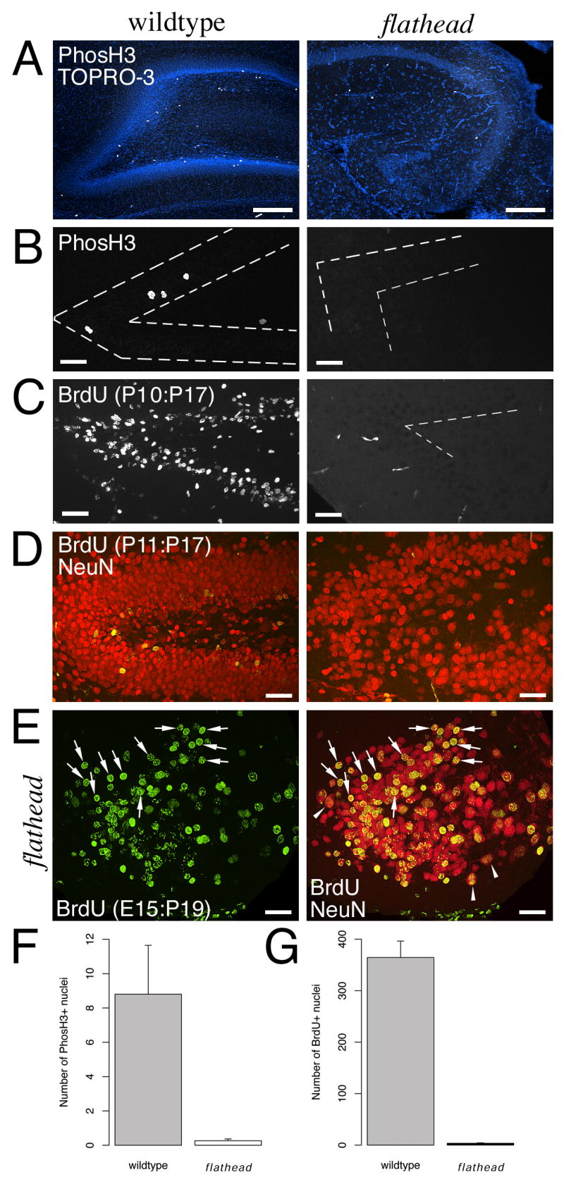
(A) Lack of PhosH3+ nuclei (white) in flathead subgranular zone (SGZ) at P16. Section is counterstained with the nuclear label TO-PRO-3 (blue). (B) Lack of PhosH3+ nuclei in flathead SGZ at P22. Dashed lines outline the granule cell layer. (C) Decreased numbers of BrdU+ cells in the postnatal flathead dentate gyrus. The images are from P17 wildtype and flathead animals that received BrdU injections at P10. The few BrdU positive objects next to the dentate gyrus in this image are blood vessels. (D) Lack of newly generated granule neurons in the flathead dentate gyrus. BrdU is green and NeuN is red. (E) Dentate granule neurons in flathead incorporate BrdU at E14-E15 (arrows). Image is from a P19 flathead that received BrdU on E14 and E15. Binucleate neurons are indicated by arrowheads. (F) Quantification of PhosH3+ nuclei in the SGZ. Numbers are expressed as the mean number of PhosH3+ cells in the SGZ per section. p < 0.05. (G) Quantification of the number of BrdU+ cells in the dentate gyrus. Numbers are expressed as mean number of BrdU+ nuclei per section. p < 0.001. Scale bars: A-E, 40μm.