


Figure 8.
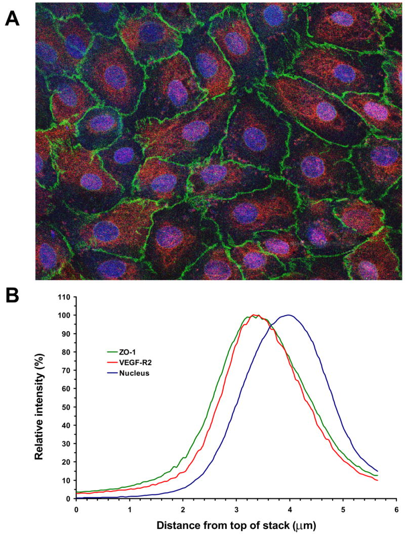
Apical localization of VEGF-R2 in confluent RPE monolayers. A, Immunohistochemical staining of the porcine primary RPE cell monolayer with mouse anti-ZO-1 and rabbit anti-VEGF-R2 as primary antibodies, and FITC-conjugated goat anti-mouse (green) and rhodamine-conjugated goat anti-rabbit (red) as secondary antibodies, respectively. The cell nuclei were visualized with DRAQ5 (blue). The image was obtained as a Z-stack on the confocal microscope. The VEGF-R2 staining was only observed in the apical regions and was absent at depths below the nuclei. The magnification was ×400. B, Color profile in the Z-stack images. The figure shows the profile for a representative region, containing a single RPE cell. Optical sectioning revealed that single color profiles represent the localization of the individual stains in the depth of the image. Analysis of color profiles revealed the close co-localization of VEGF-R2 receptor and ZO-1 protein in the apical region of the RPE cell.