


Figure 3.
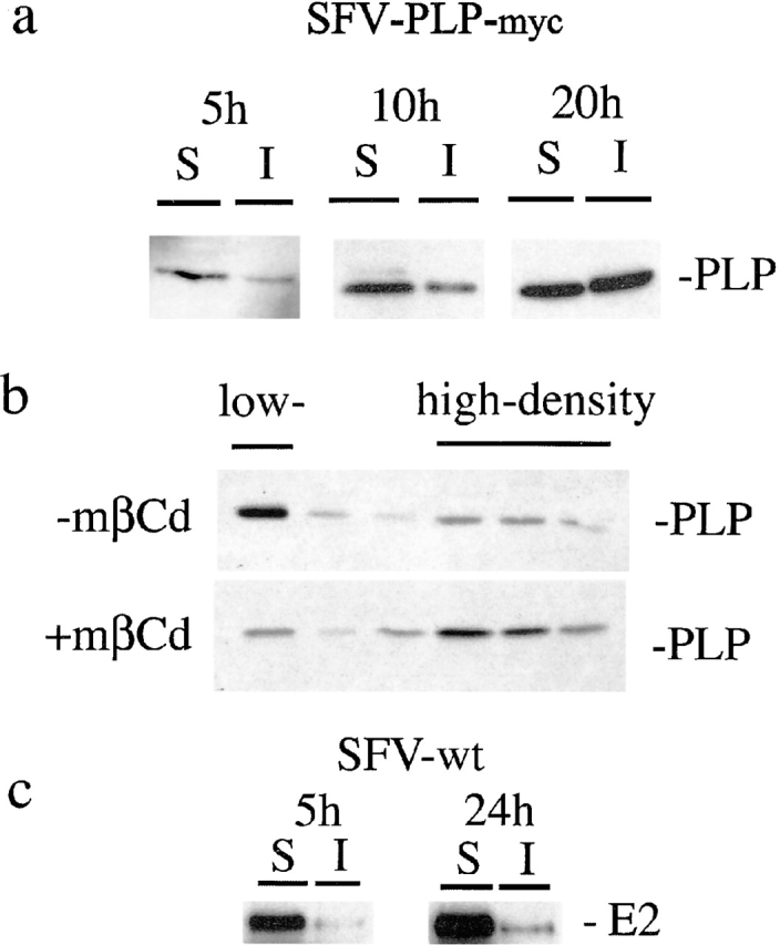
PLP associates with lipid rafts in BHK cells after increasing expression times. (a) BHK cells were infected with SFV-PLP–myc. At 5, 10, or 20 h after infection, cells were extracted with 20 mM CHAPS for 30 min at 4°C and centrifuged for 30 min at 14,000 g. Detergent-insoluble proteins in the pellet (I) and the detergent-soluble proteins in the supernatant (S) were resolved on SDS-PAGE followed by Western blotting. (b) BHK cells were infected with SFV-PLP–myc. 20 h after infection, the cells were treated with 10 mM mβCd or left untreated, extracted with CHAPS, and floated in a density gradient. Six fractions were collected from the top. The top fraction represents the low-density, CHAPS-insoluble membrane fraction, whereas the fractions of high density contain CHAPS-soluble membrane. (c) BHK cells were infected with wild-type SFV. 5 or 20 h after infection, cells were processed as described in a. The pellet (I) and supernatant (S) were analyzed by Western blotting for the viral spike protein E2.