


Figure 3.
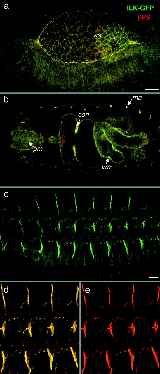
ILK colocalizes with integrins during embryonic development. (a–e) Colocalization of ILK-GFP and PS integrins. ILK-GFP is visualized by GFP fluorescence (green) and the βPS subunit is detected with a monoclonal antibody (red), with colocalization appearing yellow. Anterior is left in this and all subsequent panels. (a) Dorsolateral view of an embryo at stage 13 during dorsal closure, where ILK-GFP and integrins are concentrated at the leading edges of epidermis and the edges of the amnioserosa (as). (b) Optical horizontal section of mid-stage 16 embryo focused on the internal organs to show the localization of ILK in pharyngeal muscles (pm) and visceral mesoderm (vm), with particularly strong expression of ILK seen here at the first midgut constriction (con), and at muscle attachment sites (ma). (c) Lateral view of a late stage 16 embryo showing ILK-GFP localization at muscle attachment sites. (d and e) Same embryo at higher magnification to show the tight colocalization of ILK with integrins (d) and the integrin staining alone (e). Bars, 20 μm.