


Figure 3.
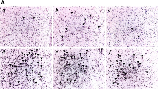

Uncontrolled encephalitis and leukocyte infiltration in the brains of 5-LO−/− mice chronically infected with T. gondii. (A) Frozen sections of brains of wild-type controls (a, b, and c) and 5-LO−/− (d, e, and f) mice infected for 25 d with T. gondii stained with anti-CD4 (a and d), anti-CD8 (b and e), or anti-CCR5 (c and f). Representative micrographs (×200) from at least three animals per group are presented. Arrows indicate reactive cells in the section. The mean ± SD of the total counts of (B) CD3+ CD4+, CD3+ CD8+ cells or of the (C) CD11c+ CD11blo, CD11c+ CD11bhi cells and the intensity of the (D) latter cells obtained from brain homogenates (n = 3) of the same experimental groups is also shown. *, statistically significant (P < 0.05) differences between group means.