


Figure 7.
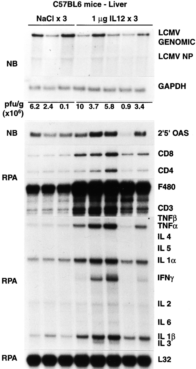
Intrahepatic LCMV, T cell marker, and cytokine gene expression after IL-12 administration. Age- and serum LCMV titer–matched C57BL/6 mice were injected intraperitoneally either with saline or with IL-12 (1 μg/d/mouse) and killed on day 3 after injection, as indicated. Northern blot (NB) analysis (top panel) was performed with 20 μg of total liver RNA from two representative mice per group. The membrane was hybridized with 32P-labeled LCMV-, 2′5′ OAS– (a marker of IFN-α/β induction), and GAPDH-specific DNA probes. Total hepatic RNA (10 μg) from the same mice was also analyzed by RNase protection assay (RPA) for the expression of various cytokine transcripts, and for the expression of CD3, CD4, CD8, and F480, as indicated. The mRNA encoding the ribosomal protein L32 was used to normalize the amount of RNA loaded in each lane. The results of the plaque assay (PFU per gram) of individual liver tissues are also indicated.