


Figure 5.
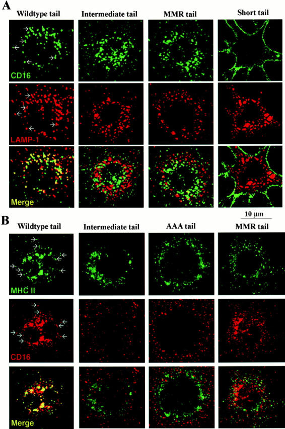
Typical distribution of CD16 chimeric receptors in DCEK.ICAM.Hi7 cells in the absence (shown here) or presence of ligand, 10 μg/ml aggHuIgG. (A) CD16 and LAMP-1 double labeling. Cells on tissue culture chamber slides were fixed and stained for CD16 (green, FITC-labeled anti–mouse Ig) and LAMP-1 (red, Texas red–labeled anti–rat Ig). Colocalization in discrete vesicles in WT transfected cells is indicated by arrows, and for all the transfectants is displayed in yellow (bottom row). (B) CD16 and MHC II double labeling. Cells grown on tissue chamber slides were fixed and stained for CD16 (red) or MHC II (green) with species-specific secondary reagents. Slides were analyzed by confocal laser scan microscopy. Examples of colocalization in single vesicles in WT transfected cells are indicated by arrows and displayed in yellow (bottom row).