


Figure 7.
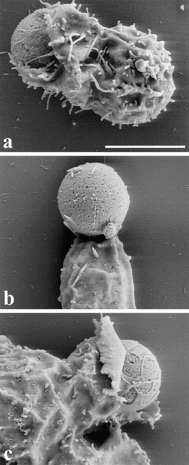
Scanning EM analysis of the interaction between Jurkat T cell and anti-CD3–coated beads. Untransfected Jurkat T cells (a), T cells transfected with GFP-ActA repeats (b), and T cells transfected with GFP-ActA repeats F > A (c) were incubated with anti-CD3–coated beads, fixed, and processed for scanning EM. Jurkat T cells, which expressed GFP-ActA repeats, formed a very loose contact with the bead. In contrast, control T cells or cells which expressed the mutated GFP-ActA repeats extended sheet-like protrusions that almost completely surrounded the beads. Bar, 5 μm.