


Figure 3.
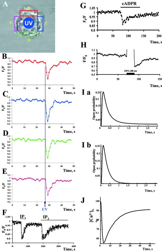
Effects of rapid applications of IP3 and cADPR to isolated nuclei loaded with Mag-Fura Red™. (A) Arrangement for experiments with uncaging of IP3 inside the nucleus (see Materials and methods). The colored boxes correspond to the colored traces in B–E, representing the Ca2+ concentrations in the indicated parts of the nuclear envelope, during uncaging of IP3 in the center of the nucleus (UV). (F) Effects of ionophoretic IP3 application. (G) Effect of ionophoretic cADPR application. (H) Bleaching-recovery experiment in isolated nucleus. Bleaching by laser light at 488 nm caused marked reduction in fluorescence intensity in affected area followed by fluorescence recovery due to diffusion from nonbleached areas. (I) Simulated time course of the open probability of (a) type 2 and (b) type 3 IP3 receptors in response to 20 μM IP3. (J) Simulation of the release and reuptake of Ca2+ in the nuclear envelope in response to IP3 stimulation. [IP3] = 20/(1 + e(−(t−10)/2)) (μM) in this simulation, to model the gradual increase of IP3 by perfusion (for further details see supplemental information, available at http://www.jcb.org/cgi/content/full/jcb.200306134/DC1).