


Figure 4.
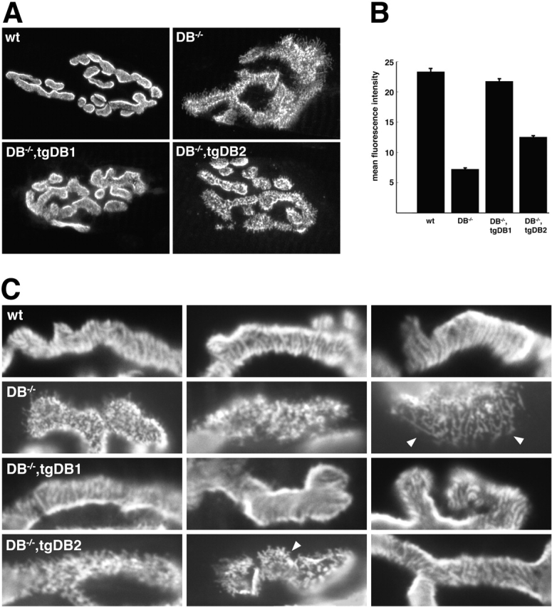
αDB1 is better able than αDB2 to restore normal AChR density and distribution to αDB −/− mice. (A) Longitudinal sections of wild-type, αDB−/−, αDB−/−,tgDB1, and αDB−/−,tgDB2 muscle stained with rBTX. In wildtype synapses, AChRs form multiple branches with smooth borders. Within branches, AChRs are evenly distributed or faintly striated. In αDB−/− synapses, AChRs are patchily distributed with fragmented branch borders and radiating spicules. In αDB−/−,tgDB1 synapses, AChR pattern is essentially normal. In the majority of αDB−/−,tgDB2 synapses, AChRs are patchily distributed, but spicules of AChRs at branch borders are shorter than those of mutants. (B) Synaptic density (± SE) of AChRs in each of the four genotypes as assessed by their mean fluorescence intensity after incubation with a saturating dose of rBTX. AChR density in αDB−/− synapses (n = 49) is ∼30% of wild-type (n = 18). At αDB−/−,tgDB1 synapses (n = 49), AChR density recovers to control levels, whereas in αDB−/−,tgDB2 synapses (n = 19) density remains intermediate between wild-type and αDB−/−. (C) High power images of branches from synapses such as those shown in A to highlight differences in AChR patterning. Spicules are indicated by arrowheads.