


Figure 6.
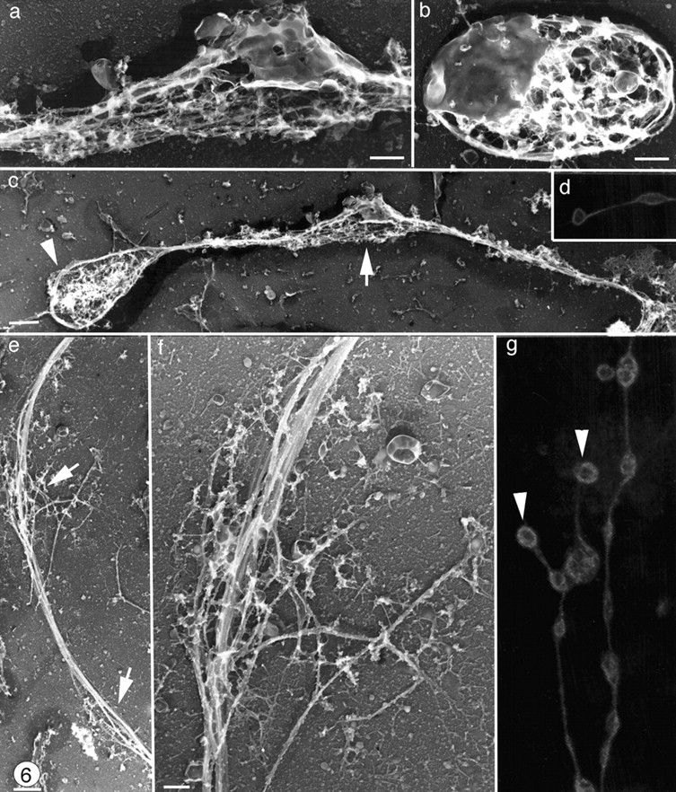
Organization of microtubules along the shaft of proplatelets. Swellings along the proplatelet (a) contain detergent-insoluble membrane and vesicles that are similar to those found in released platelets (b) in these samples. (c) Low magnification electron micrograph showing the microtubules bundles lining the shaft of a typical proplatelet. Although proplatelet ends (arrowhead) have distinct microtubule coils, microtubules observed in swellings along the shaft (arrow) are not coiled. (d and g) Proplatelets stained for tubulin by immunofluorescence and photographed on a confocal microscope. Proplatelets show periodic segments connected by thin cytoplasmic bridges, but only the ends (arrowheads) have microtubule bundles arranged into teardrop-shaped loops. (e and f) Higher magnification electron micrograph of a swelling along a proplatelet shaft showing that these are points where the microtubule bundle separates for a short distance (arrows) but does not form a loop. Bars: (b and f) 0.5 μm; (c and e) 1 μm.