


Figure 7.

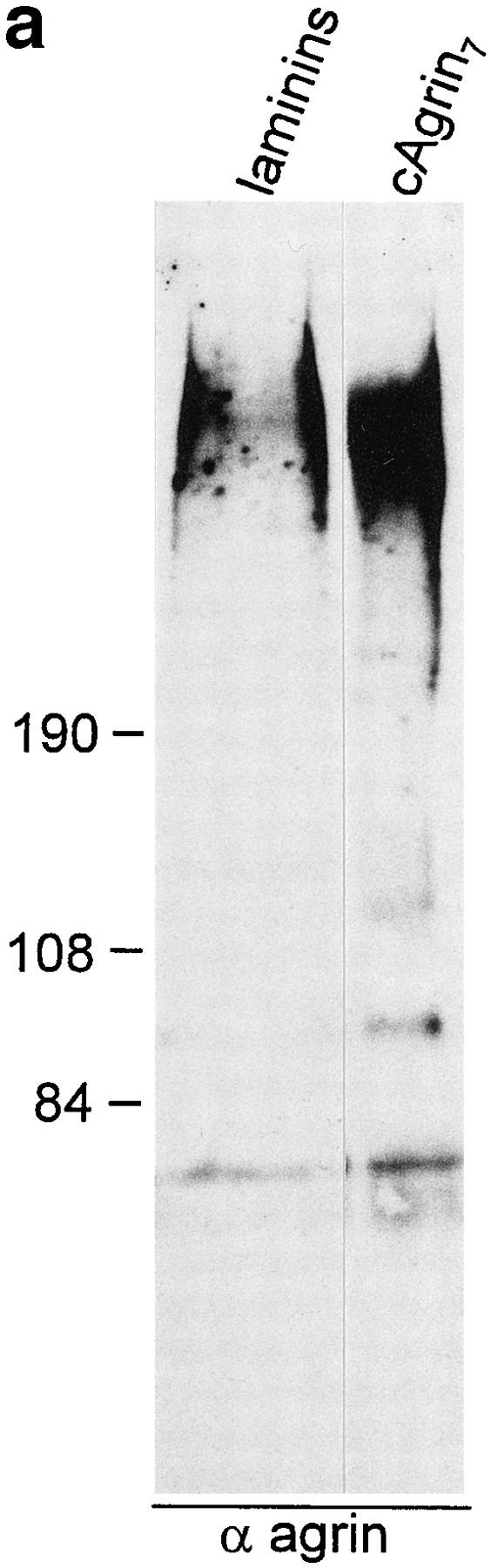
Agrin binds to laminin isoforms expressed in muscle. (a) Immunoblot using antiagrin antibodies of laminin isoforms purified from EDTA extracts of adult chick heart by immunoaffinity column with the anti-γ1 subunit–specific mAb 11B7 (Brandenberger and Chiquet, 1995). Agrin-like protein with the same apparent molecular mass as cAgrin7 is detected in the laminin preparation. Since no α-dystroglycan was detected in the same laminin preparation (data not shown), the copurification of agrin suggests that laminins are associated with agrin in muscle tissue. (b and c) Agrin binds directly to purified chick laminin-2 and -4. Equal amounts of the two laminins were immobilized on microtiter plates and incubated either with 5 nM of iodinated cAgrin7 (b) or cN257Fc (c). Values given are the result of one representative experiment and represent the mean ± SD of three measurements where the background (BSAcoated wells) has been subtracted. Background values were 163 ± 24 cpm in b and 75 ± 11 cpm in c. The binding of cAgrin7 to laminin-4 is fivefold stronger than to laminin-2 (b) and a similar difference in the binding (fourfold) is observed with cN257Fc (c). Note that the difference in the counts measured with 125I-cAgrin7 and 125I-cN257Fc is due to different iodination efficiencies.