


Figure 7.
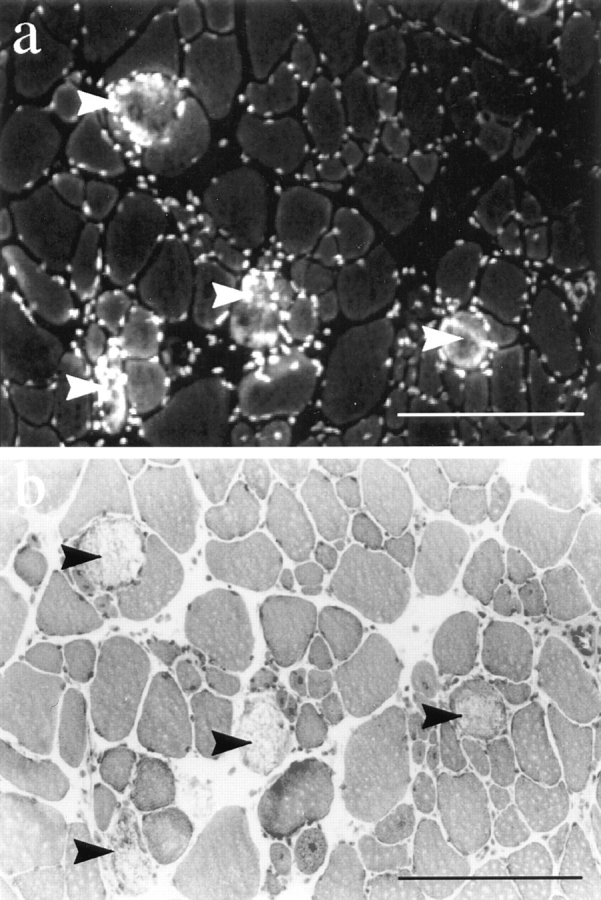
EBD staining (a) and H&E staining (b) on 7-μm cryosections from a 7-wk-old dy/dy diaphragm. The few EBD-positive fibers in dy/dy skeletal muscle always showed necrotic features in the corresponding H&E stain. The nuclei of infiltrating immune cells were also detected by the EBD. Bars, 100 μm.