


Figure 5.
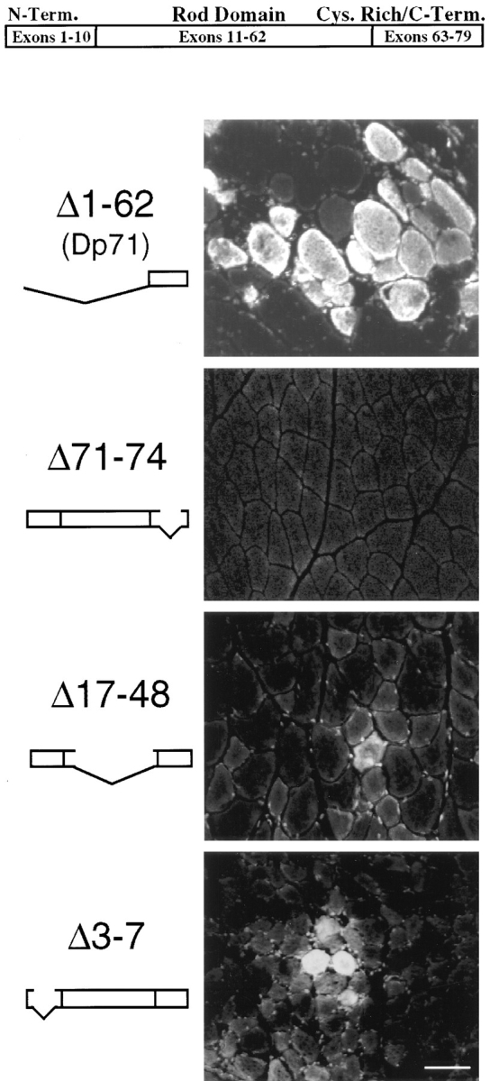
EBD staining on 7-μm cryosection of skeletal muscle from four transgenic/mdx mice. The top portion of the picture shows a model of the normal dystrophin gene. The femoral quadriceps muscle of the Dp71 mouse (20 wk old) revealed the same amount of fiber damage in the EBD assay as the original mdx mutant. Mice with a deletion of dystrophin exons 71–74 (17 wk old) did not show significantly more dye uptake into the femoral quadriceps muscle than seen in control animals. The Δ17-48 transgenic/mdx mouse (9 wk old), which has a deletion in the rod domain of dystrophin, showed few dye-positive fibers in the quadriceps femoris muscle. The Δ3-7 transgenic/mdx mouse (10 wk old), which has a deletion in the amino-terminal actin-binding domain of dystrophin, showed dye-positive fibers in the diaphragm (Δ3-7), as well as in the quadriceps femoris muscle. Bar, 50 μm.