


Figure 5. Lens development: the earliest stages.
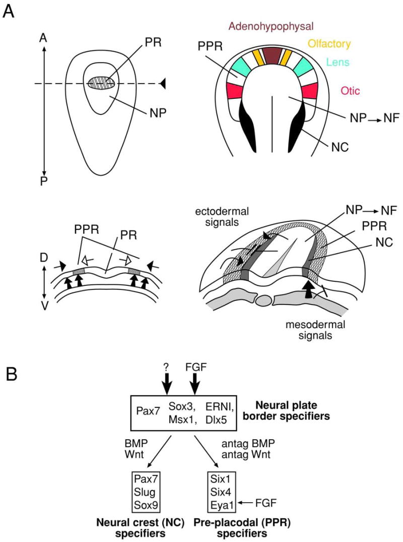
(A) Neural plate stage (left) and transition from neural plate to neural folds (right). The position of future placodes in the PPR is indicated by colors: adenohypophysal (brown), olfactory (yellow), lens (green) and otic (red). The signals that are thought to form the PPR are shown by arrows. The candidate signaling pathways/molecules are discussed in text. Adopted from (Litsiou et al., 2005). Neural crest, NC; neural folds, NF; neural plate, NP; pre-placodal region, PPR; prospective retina, PR.