


Figure 3.
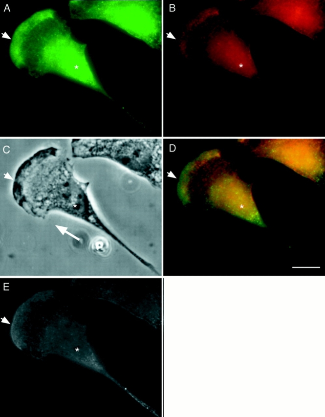
Bimodal distribution of P-myosin in a motile fibroblast of REF-2A. A motile REF-2A cell was double labeled with pp2b (A), and the monoclonal myosin antibody (B). (C) Phase contrast image. (D) Merged image (green, pp2b; red, anti–heavy chain antibody). (E) Ratio image of A/B. As shown in the merged image of D, both anterior (arrowhead) and posterior (the tail in particular) regions of the cell are greenish whereas the middle area is reddish, indicating that P-myosin is enriched in both anterior and posterior regions. The ratio image of E supports this notion. Asterisk, nucleus. A large white arrow in C shows the direction of cell locomotion. Bar, 10 μm.