


Figure 5.
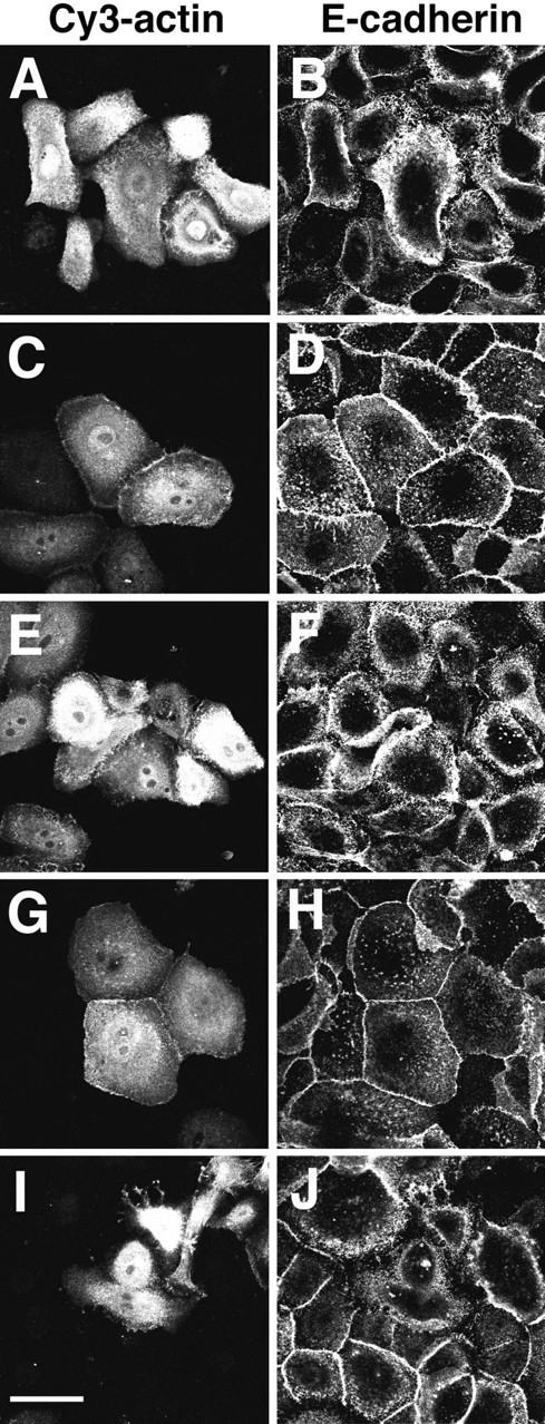
Cy3-actin is rapidly concentrated at sites of cadherinmediated adhesion upon induction of stable cell–cell contacts. Keratinocytes grown in low calcium medium were microinjected with Cy3-actin and either maintained in the same medium (A and B) or transferred to standard medium for 20 min (C–J). Control IgG (G and H) or blocking-functional antibodies against cadherins (E and F) were incubated with the cells before the microinjection of labeled actin. In other experiments, Cy3-actin was coinjected with C3 (I and J) before induction of cell–cell contacts for 20 min. The same results were obtained by coinjection of Cy3actin with N17Rac (data not shown). Microinjected cells were identified (A, C, E, G, and I), and immunostaining for E-cadherin was performed (B, D, F, H, and J). Bar, 50 μm.