


Figure 5. Representative images, histological sections and example RNA in situ hybridisation expression patterns demonstrating structures within the embryonic and adult pelvis and ureter.
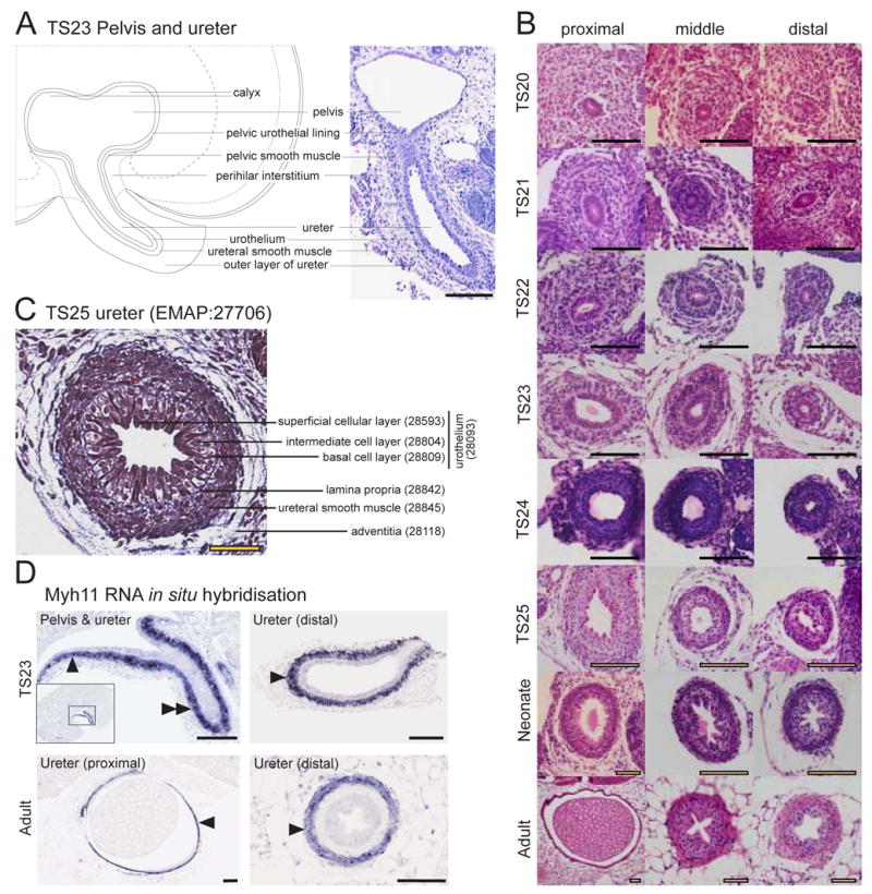
A. Low resolution diagrammatic representation (left) and histological section (right, haematoxylin and eosin) of a TS23 metanephros identifying structures around the pelvis and pelvic-ureter junction. Scale bar = 100μm. B. Temporal panel of transverse sections of the ureter at differing distances from the metanephros (proximal, middle and distal) demonstrating the histology of the developing ureter from TS20 to adult (haematoxylin and eosin). Scale bar = 100μm. C. Annotated section through a TS25 ureter middle distance between the metanephros and bladder demonstrating the cellular layers (Massons Trichrome, which stains the lamina propria blue). Scale bar = 50μm. D. Myh11 RNA in situ hybridisation of TS23 and adult ureter. TS23 Pelvis & ureter: Myh11 showed specific expression in the pelvic smooth muscle (single arrowhead, EMAP:28121) and ureteral smooth muscle layer of the ureter (double arrowhead, EMAP:28812). Inset image shows the entire metanephros section and outlines the region enlarged in the main image. TS23 Ureter (distal): Myh11 expression in the ureteral smooth muscle layer (single arrowhead, EMAP:28812) of the ureter located close to the primitive bladder. Adult Ureter (proximal): Myh11 expression in the ureteral smooth muscle layer of the ureter (arrowhead, EMAP:29488) located close to the metanephros. Adult Ureter (distal): Myh11 expression in the ureteral smooth muscle layer of the ureter (arrowhead, EMAP:29488) located close to the bladder. Scale bar = 200μm. Images courtesy of Eleanor Mitchell, Kylie Georgas, Hansheng Chiu and Bree Rumballe. Line diagram courtesy of Kylie Georgas.