



SUMMARY
Two bilaterally symmetric eyes arise from the anterior neural plate in vertebrate embryos. An interesting question is whether both eyes share a common developmental origin or they originate separately. We report here that the expression pattern of a new gene ET reveals that there is a single retina field which resolves into two separate primordia, a suggestion supported by the expression pattern of the Xenopus Pax-6 gene. Lineage tracing experiments demonstrate that retina field resolution is not due to migration of cells in the median region to the lateral parts of the field. Removal of the prechordal mesoderm led to formation of a single retina both in chick embryos and in Xenopus explants. Transplantation experiments in chick embryos indicate that the prechordal plate is able to suppress Pax-6 expression. Our results provide direct evidence for the existence of a single retina field, indicate that the retina field is resolved by suppression of retina formation in the median region of the field, and demonstrate that the prechordal plate plays a primary signaling role in retina field resolution.
Keywords: bilateral symmetry, retina field, prechordal mesoderm, eye development
INTRODUCTION
Formation of bilaterally symmetric and asymmetric structures have been studied in vertebrates since the beginning of experimental embryology. It is known that several strategies are used to form these structures in the embryo. Most of the structures on both sides of an animal, such as limbs, emerge independently from separate primordia on each side of the embryo. Formation of a single heart, on the other hand, results from the fusion of two originally separate primordia (Copenhaver, 1926; Jacobs and Fraser, 1994). Here we address the question how two symmetric eyes form in the anterior neural plate.
Interest in how two eyes form in normal embryogenesis has, in part, been motivated by attempts to understand developmental origins of cyclopean eyes in abnormal situations. A single eye or two fused eyes exist in some human fetuses suffering from holoprosencephaly (Cohen, 1989; Sperber et al. 1987; Kallen et al. 1992; Muenke et al., 1994), a developmental abnormality occurring in about 1 out of 16,000 live births and 1 in 250 terminated conceptuses (Muenke et al., 1994; Cohen, 1989). Cyclopean eyes in various species have been described with reasonable accuracy since the sixteenth century. Two mechanistic explanations were offered in the last century. Speer (1819) and Meckel (1826) proposed that cyclopia resulted from the fusion of two originally separate eyes, whereas Huschke (1832) proposed that there was a single vesicle which later became two optic vesicles in normal embryos and that cyclopean eyes resulted from a failure in separating the single vesicle (reviewed by Adelmann, 1936a). The debate continued into this century. Spemann believed in the existence of two separate optic anlagen in the early neural plate (Fig. 1A; Spemann, 1938; Lewis, 1907; Stockard, 1913b; Adelmann, 1936a). His view of separate eye anlagen in normal embryos led to his interpretation of cyclopean eyes as resulting from the fusion of two eye anlagen. His opinions were shared by King (1905) and Lewis (1907, 1909). In contrast, Stockard (1907a,b, 1908a,b, 1909a,b, 1910a,b, 1913a,b, 1914), argued that there was a single eye anlage in the median region of anterior neural plate which spread laterally and separated eventually into two eyes (Fig. 1B; Stockard, 1913a,b). LePlat (1919) came to a conclusion similar to that of Stockard except that he viewed the anlage as ‘optico-ocular’ apparatus including both the eyes and the optic chiasma (Fig. 1C). A series of experiments by Adelmann determining the developmental potential of ectodermal regions in amphibian embryos revealed that both the lateral and median portions of the anterior neural plate could give rise to eyes (Adelmann, 1929a,b, 1930, 1934, 1936), suggesting the existence of an equipotential eye field (Adelmann 1929a,b; Fig. 1D). However, since early embryos are known to have regulative ability, Adelmann cautiously pointed out that it was not clear whether eye formation by the median region isolated from its original surroundings reflects its potential in normal development (1929b). In fact, prosencephalon areas anterior and posterior to the eye field could also form eyes when isolated from their normal neighbors (Corner, 1963 and 1966; Boterenbrood, 1970). These observations have been interpreted as revealing a forebrain field which includes the telencephalon, the eyes and other regions of the diencephalon (Fig. 1E; Corner, 1963, 1966; Boterenbrood, 1970; Nieuwkoop et al., 1985), making it unclear whether there is a distinct eye field or that Adelmann’s results simply showed part of properties of the entire forebrain field. The classic studies have been largely forgotten (Hamburger, 1988, p62; Nieuwkoop et al., 1985) and the hypothesis of an eye field remains controversial.
Fig. 1.
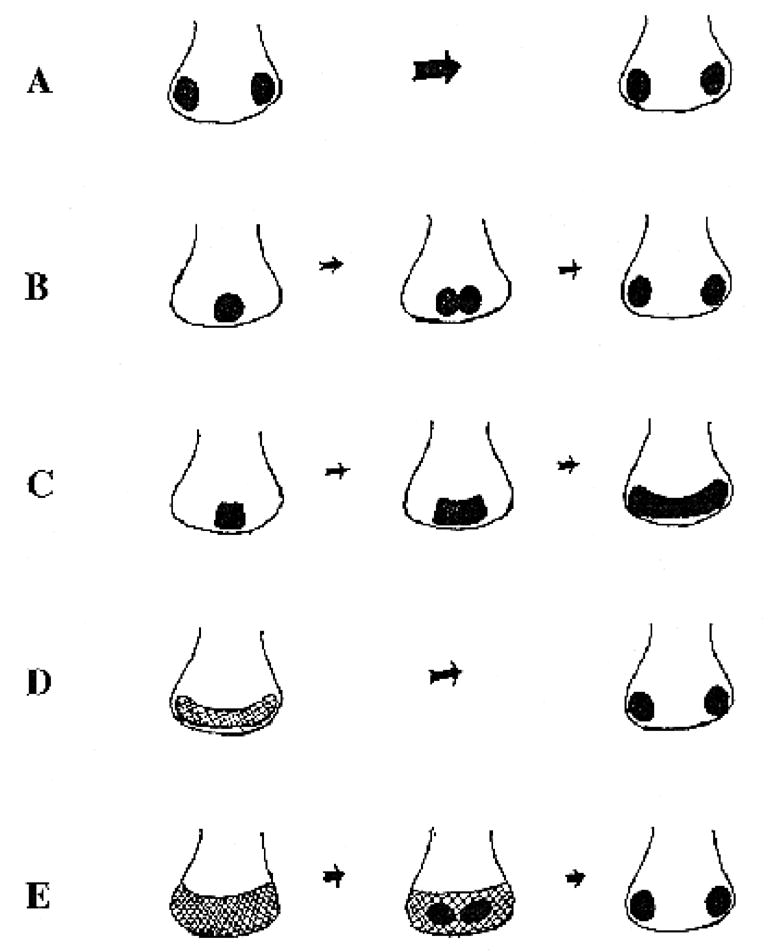
Models for retina primordium formation in vertebrate embryos. The outline illustrates the boundary of the neural plate and the filled circles represent retina primordia. (A) The two separate retina primordia model. (B) A diagram of Stockard’s model of how a single primordium spreads and separates into two retinae. Some of Stockard’s results were directly contradicted by later studies of Adelmann (1929a,b). (C) This is essentially the same as Figs 1, 2 and 4 of LePlat (1919). (D) A schematic representation of Adelmann’s model. It was not determined whether midline cells could migrate laterally. (E) An illustration of the forebrain field model. Eyes are presumably the default fate of this field (Corner, 1966; Boterenbrood, 1970).
We have been working on a new family of developmental regulators, the T domain proteins. T domain is the DNA binding domain of this new family of transcription factors (Herrmann and Kispert, 1994; Kispert et al., 1994, 1995; Kispert and Hermann, 1993) and T domain proteins have been found in both vertebrates and invertebrates (Pflugfelder et al., 1992a,b; Kispert et al., 1994; Yasuo and Satoh, 1994; Bollag et al., 1994; Bulfone et al., 1995). The prototype of the T family, the mouse T locus gene and its homologs, brachyury (Xbra) in Xenopus and no tail (ntl) in zebrafish, are essential for posterior mesoderm and notochord formation (Chesley, 1935; Grüneberg, 1958; Bennet, 1975; Hermann et al., 1990; Smith et al., 1991; Cunliffe and Smith, 1992; Schulte-Merker et al., 1992, 1994; Halpern et al., 1993). Injection of a mutant form of Xbra led to formation of the retina and the cement gland, suggesting that other T domain proteins might also exist in early Xenopus embryos (Rao, 1994).
We have now identified several genes encoding new Xenopus T domain proteins. One of them, named ET, was found to be expressed in the primordia for the retina and the cement gland. Interestingly, its early expression was in a continuous band in the anterior neural plate which later resolved into two retina primordia. To confirm that this pattern reveals a retina morphogenetic field and its resolution, we isolated and characterized the expression pattern of a Xenopus homolog of the Pax-6 gene, which is known to be important for eye formation in vertebrates and invertebrates. Xenopus Pax-6 expression pattern indeed supports the location and resolution of a retina field. Fate-mapping experiments indicate that retina field resolution is achieved by suppression of retina formation in the medial region, rather than by migration of retina forming cells into the lateral regions of the retina field. Results from experiments with Xenopus explants and chick embryos indicate that a primary signal responsible for resolving the retina field originates from the midline region of the prechordal mesoderm, the prechordal plate.
MATERIALS AND METHODS
Isolation of cDNAs
The sequences of the primers for T domain protein genes are: (GGI MGI MGI ATG TTY CCI GT) coding for GRRMPFP as the upstream primer, and (TAI GCI GTI ACI GCD ATR AA) coding for FIAVTAY as the downstream primer. The conditions for polymerase chain reactions (PCR) were: 1 cycle of 94°C, 3 minutes (min.), 53°C, 1 min. and 72°C, 2 min., followed by 36 cycles of 94°C, 45 sec., 53°C, 1 min. and 72°C, 2 min. A band of about 450 bp was obtained and subcloned into pBluescript SK. About 170 individual cDNAs were isolated and more than 90 have been sequenced. 28 of them encode 7 distinct T domain containing proteins. The sequence predicted from one of the PCR fragments corresponding to the gene named ET is shown in Fig. 2.
Fig. 2.
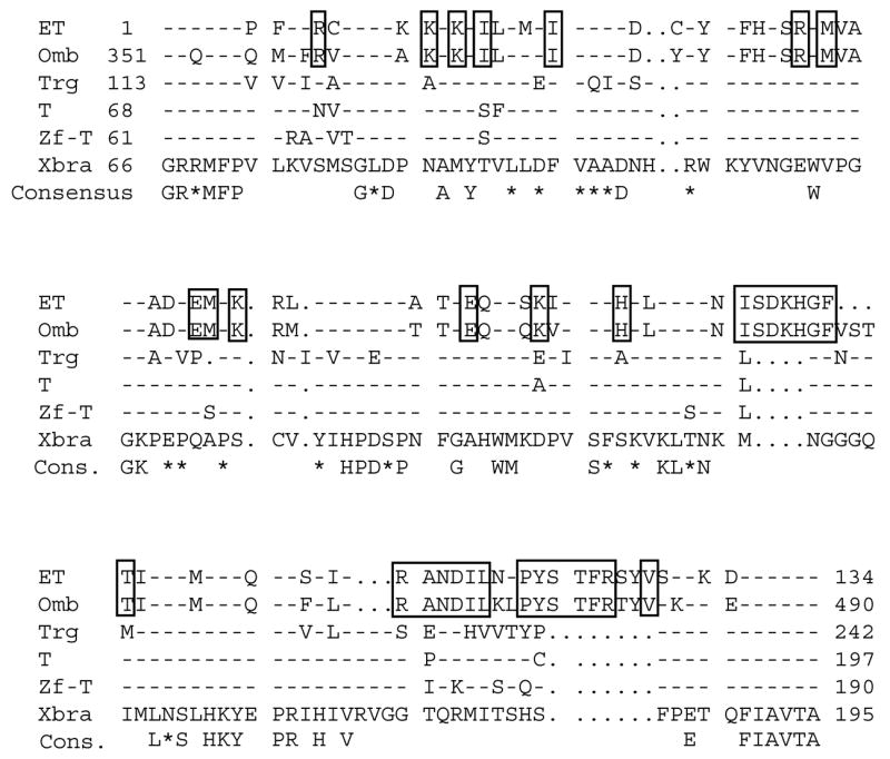
Alignment of the predicted primary sequence within the T domain of ET with those of other T domain proteins. A dash (-) indicates identity to Xbra. A period (.) indicates a gap. Residues shown in the consensus line are those identical in all known T domains while a star (*) indicates a highly but not absolutely conserved residue. Boxed residues are conserved only between ET and Omb. Omb is the predicted product of the Drosophila gene optomotor blind (Pflugfelderet al., 1992). Trg is that of the Drosophila T related gene involved in hindgut development (Kispert et al., 1994). T, Zf-T (or Ntl) and Xbra are the mouse, zebrafish and Xenopus T proteins respectively (Hermann et al., 1990; Smith et al., 1991; Schulte-Merker et al., 1992). Numbers for the ET sequence indicate positions predicted from the PCR fragment while numbers for known T domain proteins are positions in corresponding full-length proteins.
The sequences of the primers for Pax-6 are: (GIC CIC/T TIC CIG AT/CA/T G/CIA C) as the upstream primer, and (GGI AA/GA/G TCA/G/T ATC/T TTI GCI GC) as the downstream primer. The upstream primer was designed for PLPDST in the paired domain and the downstream primer was based on AAKIDLP in the homeo domain (the underlined sequence were unique to Pax-6). Template cDNAs were made from stage 28 and 40 embryos. The PCR conditions were the same as those used for isolating genes encoding T domain proteins. A fragment of 1.2 kb was subcloned into pBluescript SK and more than 150 colonies obtained. cDNAs from 16 individual clones were analysed and 15 of them had inserts. Sequence analyses showed that 10 of them contained the same sequence (of Pax-6) and the other sequences were not similar to any sequences in the databank. A probe made from the Pax-6 PCR fragment was used to screen the stage 28 cDNA library. 10 positives clones were isolated and the longest cDNA of 2.6 kb was sequenced.
Whole-mount in situ hybridization
Whole-mount in situ hybridization of Xenopus embryos was performed essentially according to the method of Harland (1991). Briefly, embryos of different stages were removed from the vitelline membrane and fixed in MEMFA (0.1 M MOPS, pH 7.4, 2 mM EGTA, 1 mM MgSO4, 3.7% formaldehyde). Digoxigenin incorporated probes were prepared with appropriate templates and hydrolyzed. Embryos were hybridized with these probes followed by sequential washes and addition of alkaline phosphatase (AP) conjugated anti-digoxigenin antibodies. AP was detected by reaction with BCIP (5-bromo-4-chloro-3-indolyl phosphate) and NBT (nitro blue tetrazolium) or BM purple as substrates.
Xenopus embryonic explants
Anterior neural plate explants were isolated from stage 12.5 or 13 embryos. The entire prechordal mesoderm was removed from the ectoderm by hair-loops and glass needles. Ectodermal explants with or without the mesoderm were then cultured in 0.5×MMR (modified Ringer’s solution) to appropriate stages. Explants were cultured to stages 18, 19, 35 and 42 and fixed in MEMFA for in situ hybridization or morphological examination. Some stage 42 explants were hybridized with a Pax-6 probe, embedded in plastic resin (JB-4 embedding kit, Polysciences, PA) and sectioned using an ultramicrotome.
DiI labeling in Xenopus embryos
This was performed essentially according to Eagleson and Harris (1990). Small chips of DiI (1, 1′-dioctadecyl-3, 3, 3′, 3′-tetramethylindocarbocyanine) were placed into the medial region of the retina field at stages 12 and 12.5.
For fate mapping, DiI-labeled embryos were cultured to stage 25 and fixed with MEMFA. They were viewed under the microscope both with epifluorescence and with incident white light. The fluorescent and incident light images were taken of each embryo at the same position using a video camera and stored in the computer as Adope Photoshop RGB files. Imposition of the two images were carried out by copying the red layer of the fluorescent image and pasting it onto the red layer of the regular lighting image, resulting in a composite image (Fig. 6D).
Fig. 6.
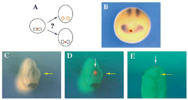
Fate mapping of midline cells in the retina field of Xenopus embryos. A to D show anterior views. (A) A diagram showing that midline cells in the retina field were labeled by DiI at stages 12 or 12.5 and followed to stage 25 to determine whether the midline cells migrate to the lateral regions. (B) The original location of DiI-labeled cells. This embryo was labeled with DiI at stage 12 and fixed at stage 12.5. DiI was photoconverted to produce the brown spot in the middle of the retina field, which was revealed by in situ hybridization with the Xenopus Pax-6 probe. (C) An image of a stage 25 embryo illuminated by incident light. A yellow arrow points to an eye. (D) A composite of the image shown in C and an image of the same embryo viewed under rhodamine optics to map the location of the fluorescent cells. A yellow arrow points to an eye. This embryo was previously labeled with DiI at stage 12.5. Results were similar with embryos labeled at stage 12. (E) A left side view of another DiI-labeled embryo. Note the absence of DiI labeled cells in the left retina. A white arrow points to DiI-labeled cells. A yellow arrow points to an eye.
To locate the original position of the DiI-labeled cells, photoconversion of DiI followed by in situ hybridization with Pax-6 was carried out (Izpisua-Belmonte et al., 1993; Saldivar et al., 1996). Briefly, embryos were labeled at stage 12 and fixed at stage 12.5 in 4% paraformaldehyde. They were rinsed twice in PBS and once in 0.1 M Tris-HCl (pH 7.4). They were equilibrated in a solution containing 1 mg/ml of DAB (3,3′-diaminobenzidine tetrahydrochlorate) in 0.1 M Tris-HCl for 30 minutes. The embryos were placed on the surface of 0.6% soft agar covered with fresh DAB solution and illuminated under a 20× objective lens using rhodamine optics. DiI was photoconverted to a brown precipitate in about 20 minutes. Embryos were transferred to 0.1 M Tris-HCl for 30 minutes and dehydrated in methanol. In situ hybridization with Pax-6 was then carried out in these embryos as described above. Pigmented embryos were eventually bleached by bright light in the presence of H2O2.
In vitro culture and microsurgery of chick embryos
Fertile white Leghorn chicken eggs (B & E Eggs, PA) were isolated and cultured as described by Sundin and Eichele (1992). Embryos were staged according to Hamburger and Hamilton (1951). Embryos were operated on and cultured with the ventral side up on 0.3% agar plates with 50% egg white. After the operations, each embryo was covered with 50 μl of Tyrode’s solution. Culture dishes were sealed with Parafilm membrane, and incubated in a humidified chamber at 38°C. 50 μl of Tyrode’s solution was added about every 12 hours. Tungsten needles prepared by electrolytic sharpening in 2 N NaOH were used for microsurgery.
In prechordal plate removal experiments, slits were made with a needle on both sides of the prechordal plate of a stage 5 embryo. The location of the prechordal region removed is diagrammed Fig. 9B and the width of the removed region is about twice that of the notochord. Whole-mount in situ hybridization of stage 6 embryos with a sonic hedgehog (shh) probe was used to verify the removal of the prechordal plate. Embryos cultured to stages 11 or 13 were examined for Pax-6 expression by in situ hybridization. Plastic sectioning of in situ hybridized embryos was carried out as previously described (Li et al., 1994).
Fig. 9.
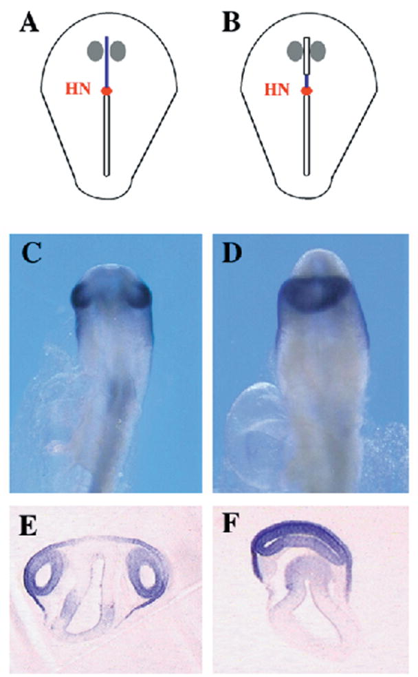
Effects of prechordal plate removal on retina formation in chick embryos. A–D are ventral views. (A) A diagram of a st. 5 chick embryo showing the location of presumptive retina primordia (indicated by two gray circles) relative to the prechordal plate (marked as a blue line) (Li et al., 1994). The red dot symbolizes Hensen’s node. (B) A diagram of the region removed from the prechordal mesoderm (indicated by a box superimposed on the blue line). (C) A st. 13 chick embryo showing Pax-6 expression in the eyes. (D) Pax-6 expression in a st. 13 chick embryo from which the prechordal plate was removed at st. 5. (E) A transverse section of a st. 13 control chick embryo after in situ hybridization with the Pax-6 probe. Ventral is up. (F) A transverse section of a st. 13 embryo which lacked the prechordal plate. The level of section is similar to that of the control embryo shown in E. Ventral is up.
In rescue experiments, mesodermal tissues from other embryos were transplanted into the host embryos from which the prechordal plate had been removed. Both the donor and the host embryos were operated on at stage 5. Positions of donor tissues are diagrammed in Fig. 10A. In order to allow proper adhesion of the transplanted tissues, host embryos were incubated with minimal amount of the culture medium for 2 hours before the addition of another 50 μl of Tyrode’s solution. Embryos with apparently proper adhesion were followed to later stages.
Fig. 10.
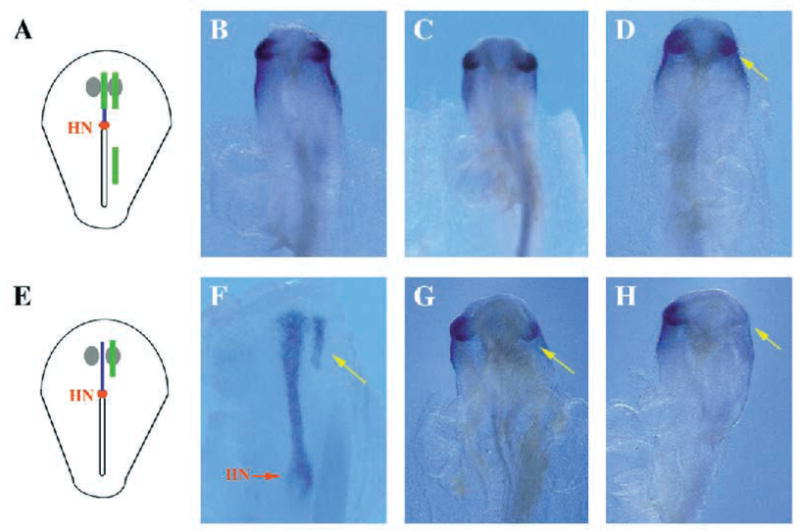
Suppression of Pax-6 expression by the prechordal plate in chick embryos. All panels show ventral views. (A) A diagram of a st. 5 chick donor embryo with the green stripes indicating the origins of the mesodermal tissues used in transplantation. The piece in the midline of the prechordal mesoderm is the prechordal plate and that lateral to the prechordal plate is the lateral prechordal mesoderm. The green stripe in the trunk region shows the origin of the lateral posterior mesodermal transplants. (B) Pax-6 expression in a wild-type st. 13 embryo. Note that Pax-6 expression is equivalent in the two eyes. (C) Pax-6 expression in a st. 13 embryo in which a piece of lateral prechordal mesoderm was inserted into the prechordal mesoderm underlying the left retina primordium at st. 5. The two eyes are equivalent. (D) Pax-6 expression is reduced in a small fraction of embryos receiving a transplanted piece of posterior lateral mesoderm (see also Table 1). This is a typical result from such transplantations with reduced Pax-6 expression in the left eye (indicated by the green arrow). (E) A diagram of the position where a transplant is placed in a host embryo (indicated by the green stripe on the left side of the embryo. (F) Shh expression in a st. 5 embryo with a transplanted piece of prechordal plate on the left side (indicated by a green arrow). A red arrow points to Hensen’s node. (G) Pax-6 expression is clearly reduced in the left eye (indicated by the green arrow) of a st. 13 embryo in which a piece of the prechordal plate was previously transplanted into a slit in the mesoderm underlying the left retina primordium at st. 5. (H) In this embryo, Pax-6 expression was more severely suppressed in the left eye (indicated by the green arrow). Strong Pax-6 suppression was observed only in embryos with prechordal plate transplants (see Table 2 legend).
Effects of mesodermal tissues on Pax-6 expression in the retina were tested by transplantations (Fig. 10A,E). A slit was made in the mesoderm underlying the left presumptive retina primordium of a stage 5 host embryo (Li et al., 1994). A piece of mesodermal tissue was isolated from a donor embryo of the same stage and placed into the slit in the host embryo. Embryos were cultured to later stages before being fixed in MEMFA.
RESULTS
Identification of ET, a gene encoding a new T domain protein in Xenopus embryos
To search for new T domain proteins, we designed degenerate primers based on conserved sequences in the T domain (Kispert et al., 1994). DNA fragments of the expected length were amplified. The amino acid sequence predicted from one of the PCR fragments was found to be similar to that of Optomotor blind (Omb; Fig. 2A), the product of a Drosophila gene essential for optic lobe development (Pflugfelder et al., 1992a,b). The gene corresponding to this PCR fragment was named ET because of its prominent expression in the eyes (see below). The predicted ET protein is also similar to that of Tbx2, a mouse T domain protein (Bollag et al., 1994). The published expression pattern of Tbx2 mRNA is not similar to that of ET (compare Fig. 6B of Bollag et al., 1994 to data shown below), making it unclear at the present whether Tbx2 is an ortholog of ET.
Pattern of ET expression suggests a single retina field
The pattern of ET expression was examined by whole-mount in situ hybridization (Harland, 1991). ET is prominently expressed in the primordia of the retina and the cement gland (Fig. 3E). ET expression in the retina primordia begins as in a single band across the midline in the anterior neural plate at stage 12.5 (indicated by the red arrow in Fig. 3A). This band of expression persists at stage 15 (Fig. 3B). Expression in the medial region of this band decreases gradually, so that it is weaker at stage 16 (Fig. 3C) and disappears by stage 18 (Fig. 3D). In late stages, ET expression in the retina is clearly localized in the dorsal part of the retina, but not in the lens or the ventral half of the retina (Fig. 3H,I).
Fig. 3.
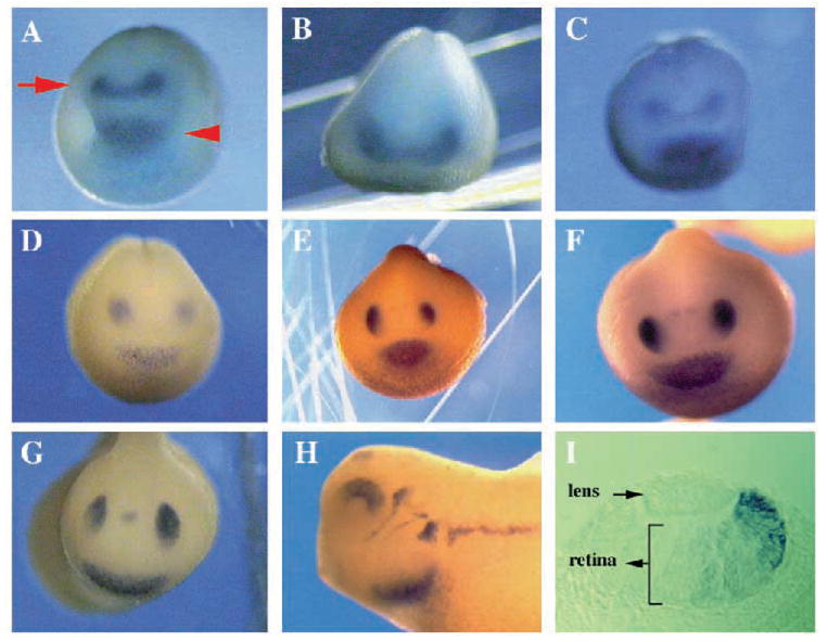
The expression pattern of ET in Xenopus embryos. A to G show anterior views and H is a lateral view. (A) A stage (st.) 12.5 embryo with a band of expression in the anterior neural plate (indicated by the arrow) and one in the primordium of the cement gland (indicated by the arrowhead). Note that the cement gland stays a single structure (A, C–G), whereas the retina band resolves into two primordia in the next few stages. (B) A st. 15 embryo showing retina field expression. The embryo was oriented to maximize visualization of retina expression and the cement gland is thus hardly visible. (C) A st. 16 embryo with ET expression weaker in the median region of the retina field than that in the lateral regions. (D) A st. 18 embryo with two distinct retina primordia. There is no ET expression in the median region. (E) A st. 19 embryo with no midline expression. (F) A st. 20 embryo. Note that weak expression of ET appears in the pineal gland primordium. The intensity of this signal increases in later stages (see G and H). (G) A st. 22 embryo with ET expression in the retina, the cement gland and the pineal gland (the dot between the two eyes). (H) A st. 26 embryo. The staining in the eye is restricted to the dorsal retina, absent from the lens and the ventral retina. Expression in the cephalic ganglia and lateral line organ also appeared. (I) A transverse section of a st. 28 embryo showing ET expression in the dorsal retina. ET is expressed in all layers of the retina, and completely absent from the lens. Dorsal is to the right.
ET is also expressed in the cement gland, an anterior epidermal structure whose formation is often associated with neural induction (Lamb et al., 1993; Hemmati-Brivanolou et al., 1994; Rao, 1994). In contrast to its expression in the retina, ET expression in the cement gland does not change its pattern from the earliest time of detection at stage 12.5 (indicated by the red arrowhead in Fig. 3A) until its disappearance around stage 26 (Fig. 3A–G). Expression in the pineal gland was detected from stage 20 onwards (Fig. 3F–H). ET is also expressed in cephalic ganglia and the lateral line organ of stage 26 embryos (Fig. 3H).
The pattern of ET expression in the anterior neural plate is consistent with the possibility of a retina field in the anterior neural plate which later resolves into two retina primordia. To ask whether ET expression as a band in the anterior neural plate reflects a general feature of genes expressed in retina primordia, we isolated another early marker for eye primordia, Pax-6 and examined its expression pattern.
Isolation of a cDNA encoding the Xenopus Pax-6 protein
Pax-6 is essential for eye formation in a wide range of species. In vertebrates such as the mouse, chicken, zebrafish and human, as well as in invertebrates such as Drosophila, Pax-6 is expressed in the eye primordia (Walther and Gruss, 1991; Krauss et al., 1991; Püschel et al., 1992; Li et al., 1994; Glaser et al., 1994, Quiring et al., 1994). Pax-6 mutations in mouse, human and Drosophila result in malformation or absence of the eyes (Hogan et al., 1988; Hill et al., 1991; Ton et al., 1991; Jordan et al., 1992; Glaser et al., 1994; Quiring et al., 1994). Ectopic expression of Drosophila Pax-6 (eyeless) in imaginal discs leads to formation of supernumerary eyes, leading to the suggestion that Pax-6 is a master regulator sufficient for initiating eye formation (Halder et al., 1995).
We isolated a cDNA fragment of Xenopus Pax-6 by PCR. This fragment allowed us to isolate cDNAs encoding an apparently full-length Xenopus Pax-6 protein (Fig. 4). Although all Pax proteins are similar within the paired and homeo domains, the primary sequences of Pax-6 proteins from all species are easily distinguishable from other Pax proteins (Quiring et al., 1994). The homeodomain of the Xenopus Pax-6 is identical to those of other vertebrate Pax-6 while all differences in the paired domain of Xenopus Pax-6 occur in regions where variations have been observed among the other Pax-6 sequences. The predicted full-length Xenopus Pax-6 product is 96% identical to mouse and human Pax-6 proteins (Fig. 4).
Fig. 4.

Comparison of Pax-6 sequence from Xenopus with those from other species. Alignment of the predicted sequences of full-length Pax-6 proteins from Xenopus, mouse and human. A dash (-) indicates identity to the Xenopus sequence. The paired domain (amino acid residues 3–133) and homeo domain (residues 209–269) are in bold.
Pax-6 expression pattern supports formation of two retina primordia from a single field
Pax-6 mRNA is first expressed at stage 12 before the completion of gastrulation and the beginning of neurulation (Fig. 5A). In the trunk region, Pax-6 is expressed in the primordium of the neural tube, beginning with two broad stripes at stage 12 (indicated by the red arrowhead in Fig. 5A). In the anterior neural plate, Pax-6 is expressed initially in a continuous band (Fig. 5A–C). Like ET, Pax-6 expression in the median region of this band is also turned off by stage 18 (Fig. 5D–G). The band of Pax-6 expression in the anterior neural plate is broader than the ET band in the anterior/posterior dimension. On each side of the midline, Pax-6 is expressed in the entire eye and a region in the forebrain (Fig. 5E,F). The precursor region for this forebrain structure is initially located in a stripe posterior to the retina field (Fig. 5B–D,G). This stripe also resolves into two bilaterally symmetric spots during neurulation (Fig. 5B–E).
Fig. 5.
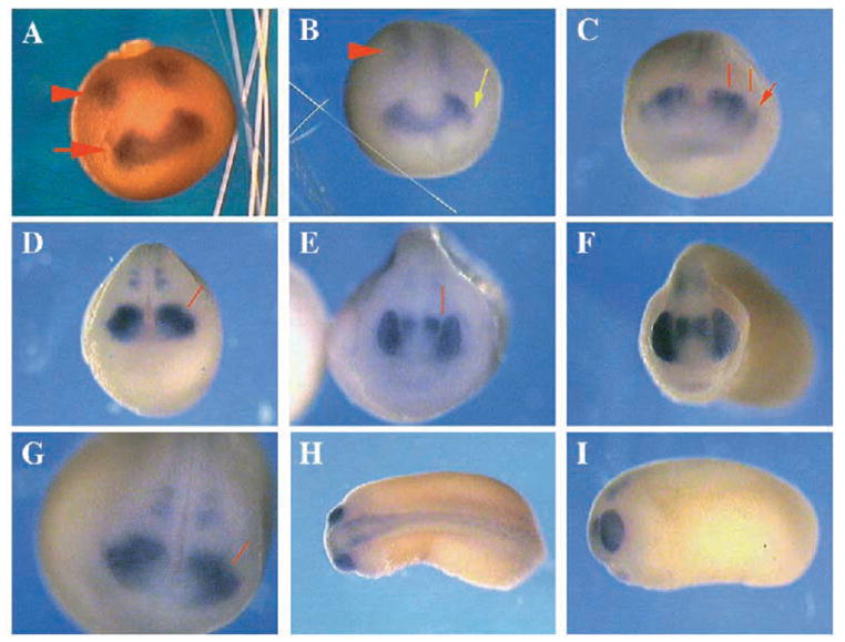
Distribution of Pax-6 mRNA in Xenopus embryos. A–G show anterior views, H is a dorsal view with the anterior end of the embryo to the left, and I is a lateral view. (A) A st. 12 embryo; note a single band of expression continuous from one side of the embryo to the other in the anterior neural plate (indicated by the red arrow) whereas there are two broad stripes of expression in the primordium of the neural tube (indicated by the red arrowhead). (B) A st. 12.5 embryo. The stripes in the trunk region are thinner and closer to each other than those at st. 12. Note the appearance of lens primordia on each side of the embryo (indicated by the green arrow). (C) A st. 16 embryo. The two red lines demarcate the borders of the retina stripe. The arrow points to a lens primordium. Pax-6 expression has three components: lens primordium, an outer semicircle of the retina field and an inner semicircle of a forebrain structure. The forebrain stripe later resolves into two spots posterior to the eyes shown in D and G, which are the two spots closer to the midline than the eyes in E and F. (D) A st. 18 embryo showing that Pax-6 expression in the median region of the anterior neural plate is turned off. The red line indicates the border between the eye primordium and the forebrain structure. Note also that the lens primordia have moved into the retina primordia to form the eye primordium by this stage (compare D to C). (E) A st. 19 embryo showing distinct Pax 6 expression in the eyes and the forebrain. (F) A stage 26 embryo. (G) Higher magnification of a st. 18 embryo. The line points to the border between the forebrain staining and the eye staining. (H) Dorsal view of a st. 24 embryo showing Pax-6 expression in the neural tube derived from the two broad stripes in the trunk region at st. 12 (the arrowhead in A). (I) A st. 25 embryo. Compare Pax-6 expression in the entire eye to ET expression in only the dorsal retina in Fig. 3H.
The Pax-6 expression pattern thus confirmed results obtained for ET in showing the presence of a continuous band in the anterior neural plate, supporting the suggestion that the expression patterns of these genes reveal a retina field which resolves into two retina primordia.
Pax-6 is also expressed in the lens, which is distinguishable from its expression in the retina field from stage 12.5 onwards (Fig. 5B,C). As early as stage 12.5, lens primordia, as revealed by Pax-6 expression, were separate on two sides of the embryo (Fig. 5B). Thus, there is so far no direct evidence for an eye field which includes both the retina and lens primordia.
Cells in the midline region of the retina field do not migrate into the lateral retina primordia
Two hypotheses can explain why retina primordia form in the lateral but not the medial regions of the retina field. One is that retina precursor cells exist in the medial region of the retina field at early stages (e.g., stages 12 and 12.5) and they migrate into the lateral regions in later stages. The alternative hypothesis is that retina formation is suppressed in the medial region of the retina field and medial cells take on fates other than that of the retina. These hypotheses can be distinguished by following the development of cells in the medial region of the retina field. The migration hypothesis predicts that some medial cells will end up in the retina, whereas the suppression hypothesis predicts that the medial cells will stay near the midline and give rise to structures other than the retina.
Previous fate mapping studies have shown that cells in the midline of the anterior neural plate of stage 15 Xenopus embryos will form the anterior pituitary gland and the supra chiasmatic nucleus (Eagleson and Harris, 1990; Eagleson et al., 1995). However, another study tracing the lineage of two-cell stage embryos suggested that retina cells from one side of the embryo could migrate to the other side (Jacobson and Hirose, 1978). Recent studies in zebrafish embryos suggest that midline cells may contribute to both retinae (Woo and Fraser, 1995). A drawback in interpreting the lineage analyses of two-cell stage Xenopus embryos is that it cannot accurately predict the fate of midline cells since the cleavage plane at the two-cell stage is variable and independent of the midline. Results from zebrafish embryos, on the other hand, may not apply to Xenopus embryos because zebrafish embryos are different from Xenopus embryos with regard to migration of early embryonic cells (Shih and Fraser, 1995). The fate of midline cells at stages 12 and 12.5 Xenopus embryos thus remains unknown.
We examined the question of cell migration by labeling midline cells in the retina field with the fluorescent dye DiI at stages 12 or 12.5 and following the fate of the labeled cells (Fig. 6A). Some of the embryos were fixed shortly after labeling in order to locate the region of DiI labeling. DiI was photoconverted to give a brown signal in these embryos. Pax-6 was used to reveal the retina field. Double labeling by DiI photoconversion and Pax-6 in situ hybridization thus allowed determination of the spatial relationship of DiI-labeled region to the retina field. DiI labeling was indeed located in the median region of the retina field (Fig. 6B).
DiI-labeled embryos were followed to stage 25 when eyes were morphologically distinguishable. Each embryo was viewed under the microscope with epifluorescence and, immediately afterwards, with incident white light. Two images from a single embryo were thus obtained through a video camera and were individually stored. Superimposition of these two images allows precise mapping of the fluorescent cells onto the whole embryo (Fig. 6D). It is clear that DiI labeled cells do not contribute to the retinae (Fig. 6D,E). These results demonstrate that cells in the midline of the retina field do not migrate into the more lateral regions to form retina precursor cells in Xenopus embryos.
Prechordal mesoderm is required for the formation of two retinae in Xenopus embryonic explants
To investigate a role for the prechordal mesoderm in the formation of two retinae (Adelmann, 1936b), we examined retina formation in explants with or without the prechordal mesoderm. Explants of the anterior neural plate were isolated from stage 12.5 or stage 13 Xenopus embryos. To ensure inclusion of the retina field in the explants, we fixed some explants immediately after isolation. They were examined by in situ hybridization with the Pax-6 probe and the retina field was found to be present in the explants (data not shown).
We then examined retina formation by allowing the explants to develop to stages 35 and 42 when pigmented retina is visible (Fig. 7A). When the anterior neural plate together with its underlying prechordal mesoderm was isolated, two retinae formed (Fig. 7B,D). In contrast, only one retina formed in explants from which the prechordal mesoderm was removed (Fig. 7C,E). Thus, the prechordal mesoderm is essential for the formation of two retinae.
Fig. 7.
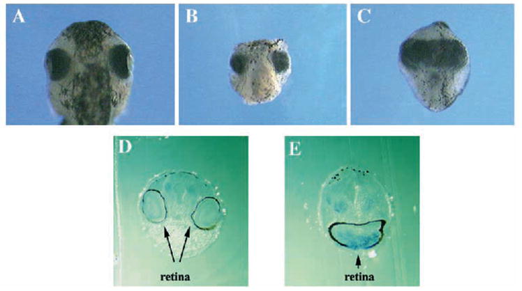
Effects of removing the prechordal mesoderm on retina formation in Xenopus embryonic explants. (A) A wild-type embryo at st. 35. (B) A st. 35 control explant of the anterior neural plate with the underlying prechordal mesoderm. (C) An anterior neural plate explant without prechordal mesoderm. The explants were isolated at st. 12.5 and cultured until st. 35. (D) A transverse section of a st. 42 control explant after in situ hybridization with a Pax-6 probe. (E) A transverse section of a st. 42 explant without prechordal mesoderm at st. 12.5 and fixed for in situ hybridization with Pax-6 at st. 42. Note the presence of Pax-6 expressing neural retina as well as the retina pigment epithelium.
Prechordal mesoderm is required for resolving the retina field
The formation of a single retina in anterior neural plate explants lacking the mesoderm could have resulted from one of the following: (1) a constant requirement of a retina inducing factor from the mesoderm for the formation of two retinae; (2) fusion of two retina primordia; or (3) failure of the single retina field to resolve. Our ability to examine retina primordia at early stages made it possible to distinguish among these different models. If the first model is true, the retina field would shrink, or in the second scenario, it would become two primordia, whereas the retina field would remain as a continuous band if the third hypothesis is correct.
We therefore examined directly the retina field with regard to the role of the prechordal mesoderm. This was carried out by hybridizing the explants with Pax-6 and ET probes. In the presence of the prechordal mesoderm, the retina field became two primordia by stage 18 (Fig. 8B,E). In the absence of the prechordal mesoderm, however, a single unresolved retina field remained (Fig. 8C,F). These results indicated that formation of a single retina in explants without the prechordal mesoderm was due to the failure of the retina field to resolve, demonstrating directly a role for the prechordal mesoderm in retina field resolution.
Fig. 8.
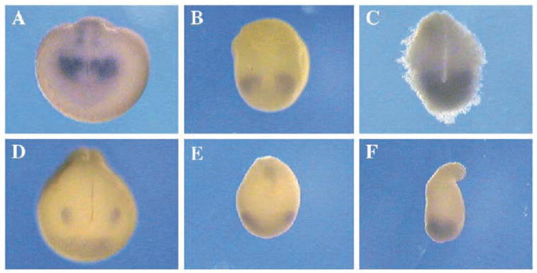
Effects of prechordal mesoderm removal on retina field resolution in Xenopus embryonic explants. (A) A control embryo hybridized with Pax-6 at st. 18. (B) Pax-6 expression in a control anterior neural plate explant with the underlying mesoderm, showing two separate primordia by st. 18. (C) Pax-6 expression in a st. 18 explant of the anterior neural plate without the underlying mesoderm. Note the retina field did not resolve into two separate primordia. (D) A control embryo hybridized with ET at st. 18. (E) ET expression in a st. 18 control explant with the prechordal mesoderm. (F) ET expression in a st. 18 explant without prechordal mesoderm.
Prechordal plate is required for the formation of two retinae in chick embryos
Explant experiments with Xenopus embryos have shown that the prechordal mesoderm is required for retina field resolution. We have carried out further experiments in chick embryos for three reasons. (1) Results discussed so far were all obtained from amphibian embryos. Their general significance remains to be tested by extending these studies to other vertebrate species. (2) Results from the explant experiments need to be confirmed by examining retina development in whole embryos. (3) The Xenopus experiments indicate retina field resolution requires the prechordal mesoderm, which is a rather large region. It is important to test the possibility that a smaller region in the prechordal mesoderm is involved in retina field resolution.
We therefore studied the consequence of removing the median region of the prechordal mesoderm, i.e., the prechordal plate, on retina formation in chick embryos. Chick embryos were operated on and cultured in albumen agar plates. The prechordal plate was removed from embryos at stage 5 (Fig. 9B) and they were cultured to stage 13 and fixed. Retinae were revealed by in situ hybridization with a chicken Pax-6 probe (Li et al., 1994). In control embryos, two retinae formed (Fig. 9C,E). In approximately 27% of embryos without the prechordal plate, a single retina, continuous from one side of the embryo to the other, formed (Fig. 9D,F). That only a fraction of embryos had cyclopia phenotype was most likely due to the difficulty in complete removal of the prechordal plate. These results thus extended the conclusions from amphibian to avian embryos and further localized the requirement for the prechordal mesoderm to its median region, the prechordal plate.
The prechordal plate could provide either a non-specific physical support or a specific signal for the formation of two retinae. To distinguish between these possibilities, mesodermal tissues from different regions were tested for their ability to rescue the cyclopia phenotype in embryos in which the prechordal plate had been removed (see Fig. 10A for a diagram of the sources of the donor tissues). The prechordal plate was able to rescue the cyclopia phenotype (Table 1), whereas mesodermal tissues taken either from the lateral parts of the prechordal region or from the lateral posterior region could not substitute for the prechordal plate (Table 1). These results indicate that the prechordal plate plays a specific role in the formation of two retinae.
Table 1.
Effects of prechordal plate removal on retina formation in chick embryos
| Number of embryos tested | Number of embryos with
|
||
|---|---|---|---|
| Treatment | 2 eyes | 1 eye | |
| a. PP removal | 51 | 37 | 14 (27.4%) |
| PP removal followed by transplantation of: | |||
| b. Prechordal plate | 39 | 39 | 0 |
| c. Lateral prechordal mesoderm | 37 | 26 | 11 (29.7%) |
| d. Lateral posterior mesoderm | 48 | 37 | 11 (22.9%) |
PP is the abbreviation of the prechordal plate. The area where the prechordal plate was removed from is shown in Fig. 9B.
The origins of tissues added back to the PP removed embryos (lateral prechordal mesoderm and lateral posterior mesoderm) were illustrated in Fig. 10A as green bars.
χ2 analyses indicate that the difference between a and b is statistically significant (P<0.01) whereas c and d are not significantly different from a (P>0.5 in both cases).
Prechordal plate suppresses Pax-6 expression in chick embryos
Our results from Xenopus explants and chick embryos suggest that the prechordal plate sends a signal to the medial region of the retina field to suppress retina formation in the medial region of the anterior neural plate. To further test this hypothesis, we carried out transplantation experiments in chick embryos to examine whether the prechordal plate could suppress the expression of Pax-6 in the retina, which should serve as a reasonable indicator for retina development because of the known role of Pax-6 in retina formation.
A slit was made in the lateral prechordal mesoderm underlying the retina primordium on the left side of a stage 5 host embryo (Li et al., 1994). A piece of the prechordal plate or mesodermal tissues from other embryonic regions was isolated from a donor embryo of the same stage, and placed into the slit in the host embryo (see Fig. 10A and E for diagrams). Embryos were then cultured to stage 13 and fixed for in situ hybridization with Pax-6.
In experiments with prechordal plate transplants, some embryos were fixed shortly after the transplantation and examined for the expression of sonic hedgehog (shh), a marker for the prechordal plate (Marti et al., 1995; Shimamura et al., 1995). Presence of a transplanted prechordal plate was confirmed by shh in situ hybridization (Fig. 10F).
In approximately 75% of embryos with transplanted prechordal plates, Pax-6-expressing areas were smaller on the side receiving the transplants (Fig. 10G, H), indicating that Pax-6 expression was suppressed. The suppression is partial in most embryos (Fig. 10G), perhaps because contact between the transplanted prechordal plate and the anterior neural plate was not as tight as that in the wild-type situation, or because the width of the region in the prechordal plate with Pax-6 suppressing activity is not large enough to cover the entire retina primordium on the left side of the chick embryo.
Neither the transplantation of a piece of lateral prechordal mesoderm, nor the making of a slit alone, affected the size of the retina on the experimental side (Fig. 10C and Table 2). Transplantation of a piece of mesoderm from the lateral posterior region of the embryo sometimes reduced the size of the retina (Fig. 10D), perhaps due to interference of retina induction after such transplantations.
Table 2.
Effects of mesodermal transplantation on Pax-6 expression in chick embryos
| Transplanted tissue | Number of embryos examined | Number of embryos with Pax-6 expression reduced on the operated side |
|---|---|---|
| a. Prechordal plate | 37 | 28 (75.7%) |
| b. Lateral prechordal mesoderm | 13 | 0 |
| c. Lateral posterior mesoderm | 34 | 9 (26.5%) |
| d. Lateral slit without any transplant | 33 | 0 |
The origins of the donor tissue are shown as green bars in Fig. 10A, and the position where the transplants were place into the host embryo is illustrated in Fig. 10E.
A typical result from prechordal plate transplantation is shown in Fig. 10G. There were 5 embryos in this group of 28 embryos with strong suppression of Pax-6 similar to that shown in Fig. 10H.
A typical result after lateral prechordal mesoderm transplantation is shown in Fig. 10C.
A typical result after lateral posterior mesoderm transplantation is shown in Fig. 10D. No complete suppression like that shown in Fig. 9H was observed after such transplantations.
χ2 analyses indicate that a is significantly different from b, c and d (P<0.01 in all three pair-wise comparisons).
The reduction of retina in embryos transplanted with the prechordal plate was more severe than that in embryos transplanted with lateral posterior mesoderm (compare Fig. 10G and D). Strong suppression like that shown in Fig. 9H was only observed after prechordal plate transplantation, but not after lateral posterior mesoderm transplantation (Table 2). Furthermore, the number of embryos with retina size reduction in those transplanted with the prechordal plate was significantly higher than that in embryos transplanted with lateral posterior mesoderm (Table 2). Taken together, these results are consistent with the suggestion that the prechordal plate can inhibit Pax-6 expression.
DISCUSSION
Our major conclusions are that there is a single retina morphogenetic field which resolves into two retina primordia, that retina field is resolved by suppression of retina formation in the median region of the field, and that the primary signal for retina field resolution comes from the prechordal plate. Mechanisms responsible for bilateral asymmetry have been investigated in recent work on the left-right axis (Brown and Wolpert, 1990; Yost, 1990, 1991, 1992, 1995; Klar, 1994; Danos and Yost, 1995; Levin et al., 1995). Our experiments have studied formation of the eyes, a model for bilaterally symmetric structures, revealing mechanisms underlying the establishment of bilateral symmetry in the forebrain.
Retina morphogenetic field and retina primordia
Our model of retina primordium formation is outlined in Fig. 11. Thus, there is a single retina field in the anterior neural plate of vertebrate embryos which becomes two primordia (Fig. 11A). This model is similar to one suggested by Adelmann who based his suggestion on the findings of retina formation in the median portion as well as the lateral portions of the anterior neural plate (Adelmann, 1929a,b). Our observations of ET and Pax-6 expression patterns in the anterior neural plate provide direct support for the existence of a single retina field in the early embryo. These results strengthened the conclusion of Adelmann in showing that the median as well as the lateral regions were indeed endowed with the potential for retina formation. Availability of the molecular markers makes it possible to locate the retina field precisely and to follow the process of its resolution into two primordia. Support for equipotentiality in the retina field also came from the experiments studying the role of the prechordal mesoderm on retina field development. Our finding of the formation of a single retina primordium in the entire field in the absence of the prechordal mesoderm (Figs 7 and 8) indicates that the entire retina field is indeed capable of forming retina. Our experiments with chick embryos further extend conclusions from studies of amphibians to other vertebrate species.
Fig. 11.
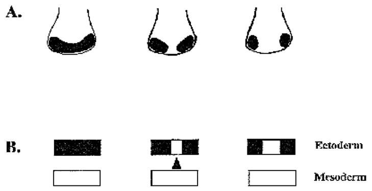
Retina primordium formation in vertebrate embryos. (A) Our model of retina field development. Dorsal views of the anterior neural plate are illustrated here. There is initially a single retina morphogenetic field in the anterior neural plate of vertebrate embryos. Retina formation is suppressed in its median region, resulting in the resolution of the retina field into two retina primordia. (B) The prechordal plate provides a primary signal for retina field resolution. These are transverse section views of the anterior neural plate and its underlying prechordal mesoderm. Experiments reported here demonstrate a role for the prechordal plate while those performed in zebrafish embryos suggest the midline of the neural plate also plays a role (Hatta et al., 1991, 1994; Hatta, 1992; Macdonald et al., 1995; Ekker et al., 1995b).
Results from our fate mapping experiments rule out the possibility that the medial region of the retina field contains retina precursor cells which later migrate into the lateral retina primordia (Fig. 6). Results from these experiments in Xenopus and ectopic transplantation of the prechordal plate in chick embryos support the suggestion that retina field resolution is achieved by suppression of retina forming potential in the median region and maintenance of retina development in the lateral regions of the retina field.
Sources of signals for retina field resolution: the prechordal plate and the midline cells of the anterior neural plate
Our experiments have addressed the role of the prechordal mesoderm in influencing the bilaterality of the retina primordia. It is shown that removal of the prechordal mesoderm could result in the formation of a single retina in the retina field. Direct examination of the retina field shows that it stays as a single field after the removal of the prechordal mesoderm (Fig. 8). Prechordal plate removal and transplantation experiments in chick embryos indicate that the prechordal plate plays a specific signaling role in inhibiting retina formation in the ectoderm (Figs 9, 10). Taken together, these results suggest that the prechordal plate provides an essential signal to resolve the retina field (Fig. 11B).
The phenotype of chick embryos removed of the prechordal plate is very similar to that of zebrafish mutants defective in the cyclops gene. Thus, fusion of Pax-6 expressing domains (Hatta et al., 1994; Macdonald et al., 1995) and formation of a single eye were observed in zebrafish cyclops mutants (Hatta et al., 1991). Cell transplantation experiments have shown that midline cells in the anterior neural plate as well as floor plate cells in the neural tube are defective in cyclops mutant embryos (Hatta et al., 1991, 1994; Hatta, 1992). Failure to form separate eyes in cyclops mutant embryos was shown to be secondary to defects of these midline cells in the anterior neural plate, suggesting that the midline cells in the ectoderm is important for the separation of the two eyes (Hatta et al., 1994).
Thus, results from amphibian and chick embryos indicate that the prechordal plate is crucial for retina field resolution while results from zebrafish embryos suggest a role for the midline cells in the anterior neural plate. This scenario is quite similar to the roles of the notochord and the floor plate in inducing ventral cell types and suppressing dorsal cell types in the neural tube (Jessell et al., 1988; van Straaten et al., 1988; Placzek et al., 1990, 1993; Yamada et al., 1991, 1993; Basler et al., 1993; Liem et al., 1995; Goulding et al., 1993).
Molecular nature of the signal for retina field resolution
Similarity of retina field resolution to dorsal/ventral patterning in the neural tube suggests that molecules responsible for neural tube patterning may also be involved in resolving the retina field. Thus, sonic hedgehog (shh), a molecule known to be important for neural tube patterning (Echelard et al., 1993; Krauss et al., 1993; Roelink et al., 1994, 1995), is a candidate signal for retina field resolution. Injection of shh mRNA into zebrafish embryos leads to overall decrease of Pax-6 expression, reduction of eyes and increased expression of Pax-2, a marker for the optic stalk (Macdonald et al. 1995; Ekker et al., 1995b). These observations, together with the finding of shh expression in the midline of the anterior neural plate (Echelard et al., 1993; Krauss et al., 1993; Roelink et al., 1994, 1995; Ekker et al., 1995a; Ericson et al., 1995), have led to the suggestion of shh as the signal in the midline of the neural plate for patterning the retina and optic stalk (Macdonald et al. 1995; Ekker et al., 1995b). Studies involving activating and inhibiting the signal transduction pathway of shh lend further support to this suggestion (Hammerschmidt et al., 1996). Observation of cyclopia in mice lacking shh proves that shh is required for the formation of two eyes. Since expression of shh is also detected in the prechordal mesoderm (Marti et al., 1995; Shimamura et al., 1995), shh may act as the prechordal plate signal for resolving the retina field. Curious findings of human fetuses with both holoprosencephaly and digit anomalies (Young and Madders, 1987; Moerman and Fryns, 1988; Shiota and Tanimura, 1988; Atkin, 1988; Cohen, 1989) could be best explained by defects in signaling pathways used both in patterning the limb bud (Riddle et al., 1993) and in determining bilaterality in the anterior neural plate.
Retina field vs. lens field
Classic literature has called the morphogenetic field in the anterior neural plate, the eye field (Adelmann, 1936b). Our examination of Pax-6 expression in Xenopus embryos shows that formation of the lens primordia is different from the retina primordia: there are two separate lens primordia as early as stage 12.5 when the retina field is still a single field (Fig. 5B). Thus, either there is no lens field or the lens field behaves differently from the retina field. In the former case, two lenses are determined from separate primordia. In the latter scenario, it is possible that at stage 12 (Fig. 5A), the lens field could not be distinguished from the retina field by examining Pax-6 expression. If this is true, then the lens field must resolve into two lens primordia by stage 12.5 (Fig. 5B). Since the retina field remains one field through stages 14, 15, 16, separation of this hypothetical lens field is accomplished earlier than separation of the retina field. Thus, whether or not a lens field exists, the mechanism underlying retina primordium formation appears to be different from that for lens primordium formation.
Bilateral symmetry in the forebrain
The Pax-6 expression pattern suggests that the bilaterality of forebrain regions other than the retina is established in a similar manner to that of the retina. Thus, a forebrain structure expressing Pax-6 was also found to be a continuous band just posterior to the retina field and this band became two spots over the same time course as the retina field resolution (Fig. 5), suggesting a mechanistic similarity in the formation of the bilaterally symmetric retina and forebrain structures. This suggestion could explain why, in cyclopean embryos, not only eyes but also other forebrain structures remain unseparated (e.g. Adelmann, 1934; Hatta et al., 1991).
Regulative changes of the forebrain primordial region observed by Corner and Boterenbrood have led to the proposal of a forebrain field (Corner, 1963, 1966; Boterenbrood, 1970; Nieuwkoop et al., 1985). This hypothesis suggests that an area larger than the retina field is equivalent in developmental potential (Corner, 1963, 1966; Boterenbrood, 1970; Nieuwkoop et al., 1985). It is not clear how the retina forms in this field, although the retina fate is supposed to be the default in this field (Corner, 1963, 1966; Boterenbrood, 1970; Nieuwkoop et al., 1985). In contrast to our data supporting the existence of a retina field, there is so far no evidence that complements the results of Corner (1963, 1966) and Boterenbrood (1970) in directly demonstrating a forebrain field.
Acknowledgments
We are grateful to V. Hamburger for discussions, M. Nonet and R. Kopan for help with microscopy, P. Bridgman for allowing us to use his ultramicrotome, G. Philips for help with plastic sectioning, C. Stern and C. Krull for advice on DiI photoconversion, O. Sundin for the chicken Pax-6 probe, R. Riddle and C. Tabin for shh cDNA, T. Fagaly, R. Kopan, J. Sanes, D. Van Essen and J. Lichtman for comments on the manuscript.
Footnotes
Note added in proof
Two papers indicating involvement of abnormality in human shh functioning as the underlying cause of a subset of holoprosencephaly have now been published.
Belloni, E., Muenke, M., Roessler, E., Traverso, G., Siegel-Bartelt, J., Frumkin, A., Mitchell, H. F., Donis-Keller, H., Helms, C., Hing, A. V., Heng, H. H. Q., Koop, B., Martindale, D., Rommens, J. M., Tsui, L.-C. and Scherer, S. W. 1996. Identification of Sonic hedgehog as a candidate gene responsible for holoprosencephaly. Nature Genet. 14, 353–356.
Roessler, E., Belloni, E., Gaudenz, K., Jay, P., Berta, P., Scherer, S. W., Tsui, L.-C. and Muenke, M. 1996. Mutations in the human Sonic hedgehog gene cause holoprosencephaly. Nature Genet. 14, 357–360.
References
- Adelmann HB. Experimental studies on the development of the eye. I The effect of the removal of median and lateral areas of the anterior end of the urodelan neural plate on the development of the eyes (Triton teniatus and Amblystoma punctatum) J Exp Zool. 1929a;54:249–290. [Google Scholar]
- Adelmann HB. Experimental studies on the development of the eye. II The eye-forming potencies of the median portions of the urodelan neural plate (Triton teniatus and Amblystoma punctatum) J Exp Zool. 1929b;54:291–317. [Google Scholar]
- Adelmann HB. Experimental studies on the development of the eye. III The effect of the substrate (‘unterlagerung’) on the heterotopic development of median and lateral strips of the anterior end of the neural plate of Amblystoma. J Exp Zool. 1930;57:223–281. [Google Scholar]
- Adelmann HB. A study of cyclopia in Amblystoma punctatum, with special reference to the mesoderm. J Exp Zool. 1934;67:217–281. [Google Scholar]
- Adelmann HB. The problem of cyclopia. Pt I. Quar Rev Biol. 1936a;11:161–182. [Google Scholar]
- Adelmann HB. The problem of cyclopia. Pt II. Quar Rev Biol. 1936b;11:284–304. [Google Scholar]
- Atkin JF. A new syndrome with cyclopia and trisomy 13 features. Am J Hum Genet. 1988;43(suppl):A36. (abstract #143) [Google Scholar]
- Basler K, Edlund T, Jessell T, Yamada T. Control of cell pattern in the neural tube: regulation of cell differentiation by dorsalin-1, a novel TGF-β family member. Cell. 1993;73:687–702. doi: 10.1016/0092-8674(93)90249-p. [DOI] [PubMed] [Google Scholar]
- Bennett D. The T-locus of the mouse. Cell. 1975;6:441–454. [Google Scholar]
- Bollag RJ, Siegfried Z, Cebra-Thomas JA, Garvey N, Davison EM, Silver LM. An ancient family of embryonically expressed mouse genes sharing a conserved protein motif with the T locus. Nature Genet. 1994;7:383–389. doi: 10.1038/ng0794-383. [DOI] [PubMed] [Google Scholar]
- Boterenbrood EC. Differentiation in small grafts of the median region of the presumptive prosencephalon. J Embryol Exp Morph. 1970;23:751–759. [PubMed] [Google Scholar]
- Brown NA, Wolpert L. The development of handedness in left/right asymmetry. Development. 1990;109:1–9. doi: 10.1242/dev.109.1.1. [DOI] [PubMed] [Google Scholar]
- Bulfone A, Smiga SM, Shimamura K, Peterson A, Puelles L, Rubenstein JLR. T-Brain-1: A homolog of Brachyury whose expression defines molecularly distinct domains within the cerebral cortex. Neuron. 1995;15:63–78. doi: 10.1016/0896-6273(95)90065-9. [DOI] [PubMed] [Google Scholar]
- Chesley P. Development of the short-tailed mutant in the house mouse. J exp Zool. 1935;70:429–435. [Google Scholar]
- Cohen MM., Jr Perspectives on holoprosencephaly: part 1. epidemiology, genetics, and syndromology. Teratol. 1989;40:211–235. doi: 10.1002/tera.1420400304. [DOI] [PubMed] [Google Scholar]
- Copenhaver WM. Experiments on the development of the heart of Amblystoma punctatum. J Exp Zool. 1926;43:321–371. [Google Scholar]
- Corner MA. Development of the brain in Xenopus laevis after removal of parts of the neural plate. J Exp Zool. 1963;153:301–311. doi: 10.1002/jez.1401530312. [DOI] [PubMed] [Google Scholar]
- Corner MA. Morphogenetic field properties of the forebrain area of the neural plate in an anuran. Experientia. 1966;22:188–189. doi: 10.1007/BF01897729. [DOI] [PubMed] [Google Scholar]
- Coulombre AJ. The eye. In: DeHaan RL, Ursprung H, editors. Organogenesis. Holt, Rinehart & Winston; New York: 1965. pp. 220–251. [Google Scholar]
- Cunliffe V, Smith JC. Ectopic mesoderm formation in Xenopus embryos caused by widespread expression of a Brachyury homologue. Nature. 1992;358:427–30. doi: 10.1038/358427a0. [DOI] [PubMed] [Google Scholar]
- Danos MC, Yost HJ. Linkage of cardiac left-right asymmetry and dorsal-anterior development in Xenopus. Development. 1995;121:1467–1474. doi: 10.1242/dev.121.5.1467. [DOI] [PubMed] [Google Scholar]
- Eagleson GW, Harris WA. Mapping of the presumptive brain regions in the neural plate of Xenopus laevis. J Neurobiol. 1990;21:427–440. doi: 10.1002/neu.480210305. [DOI] [PubMed] [Google Scholar]
- Eagleson GW, Ferreiro B, Harris WA. Fate of the anterior neural ridge and the morphogenesis of the Xenopus forebrain. J Neurobiol. 1995;28:146–158. doi: 10.1002/neu.480280203. [DOI] [PubMed] [Google Scholar]
- Echelard Y, Epstein DJ, St-Jacques B, Shen L, Mohler J, McMahon JA, McMahon AP. Sonic hedgehog, a member of a family of putative signaling molecules, is implicated in the regulation of CNS polarity. Cell. 1993;75:1417–1430. doi: 10.1016/0092-8674(93)90627-3. [DOI] [PubMed] [Google Scholar]
- Ekker SC, McGrew LL, Lai C-J, Lee JJ, von Kessler DP, Moon RT, Beachy PA. Distinct expression and shared activities of members of the hedgehog gene family of Xenopus laevis. Development. 1995a;121:2337–2347. doi: 10.1242/dev.121.8.2337. [DOI] [PubMed] [Google Scholar]
- Ekker SC, Unger AR, Greenstein P, von Kessler Porter JADP, Moon RT, Beachy PA. Patterning activities of vertebrate hedgehog proteins in the developing eye and brain. Cur Biol. 1995b;5:944–954. doi: 10.1016/s0960-9822(95)00185-0. [DOI] [PubMed] [Google Scholar]
- Ericson J, Muhr J, Placzek Lints T, Jessell TM, Edlund T. Sonic hedgehog induces the differentiation of ventral forebrain neurons: a common signal for ventral patterning within the neural tube. Cell. 1995;81:747–756. doi: 10.1016/0092-8674(95)90536-7. [DOI] [PubMed] [Google Scholar]
- Glaser T, Jepeal L, Edwards JG, Young SR, Favor J, Maas RL. Pax6 gene dosage effect in a family with congenital cataracts, aniridia, anophthalmia and central nervous system defect. Nature Genet. 1994;7:463–471. doi: 10.1038/ng0894-463. [DOI] [PubMed] [Google Scholar]
- Goulding MD, Lumsden A, Gruss P. Signals from the notochord and floor plate regulate the region-specific expression of two Pax genes in the developing spinal cord. Development. 1993;117:1001–1016. doi: 10.1242/dev.117.3.1001. [DOI] [PubMed] [Google Scholar]
- Grüneberg H. Genetical studies on the skeleton of the mouse XXIII: The development of Brachyury and Anury. J Embryol exp Morph. 1958;6:424–443. [PubMed] [Google Scholar]
- Halder G, Callaerts P, Gehring WJ. Induction of ectopic eyes by targeted expression of the eyeless gene in Drosophila. Science. 1995;267:1788–1792. doi: 10.1126/science.7892602. [DOI] [PubMed] [Google Scholar]
- Halpern ME, Ho RK, Walker C, Kimmel CB. Induction of muscle pioneers and floor plate is distinguished by the zebrafish no tail mutation. Cell. 1993;75:99–111. [PubMed] [Google Scholar]
- Hamburger V, Hamilton H. A series of normal stages in the development of the chick embryo. J Morphol. 1951;88:49–92. [PubMed] [Google Scholar]
- Hamburger V. The Heritage of Experimental Embryology: Hans Spemann and the organizer. Oxford University Press; New York: 1988. [Google Scholar]
- Hammerschmidt M, Bitgood MJ, McMahon AP. Protein kinase A is a common negative regulator of Hedgehog signaling in the vertebrate embryo. Genes Dev. 1996;10:647–658. doi: 10.1101/gad.10.6.647. [DOI] [PubMed] [Google Scholar]
- Harland RM. In situ hybridization: an improved whole-mount method for Xenopus embryos. In: Kay BK, Peng HB, editors. Methods in Cell Biology. Vol. 36. Academic Press; San Diego: 1991. pp. 685–695. [DOI] [PubMed] [Google Scholar]
- Hatta K. Role of the floor plate in axonal patterning in the zebrafish CNS. Neuron. 1992;9:629–642. doi: 10.1016/0896-6273(92)90027-b. [DOI] [PubMed] [Google Scholar]
- Hatta K, Kimmel CB, Ho RK, Walker C. The cyclops mutation blocks specification of the floor plate of the zebrafish central nervous system. Nature. 1991;350:339–341. doi: 10.1038/350339a0. [DOI] [PubMed] [Google Scholar]
- Hatta K, Püschell AW, Kimmel CB. Midline signaling in the primordium of the zebrafish anterior central nervous system. Proc Natl Acad Sci USA. 1994;91:2061–2065. doi: 10.1073/pnas.91.6.2061. [DOI] [PMC free article] [PubMed] [Google Scholar]
- Hemmati-Brivanolou A, Kelly OG, Melton DA. Follistatin, an antagonist of activin, is expressed in the Spemann organiser and displays direct neuralizing activity. Cell. 1994;77:283–295. doi: 10.1016/0092-8674(94)90320-4. [DOI] [PubMed] [Google Scholar]
- Herrmann BG, Labeit S, Poustka A, King TR, Lehrach H. Cloning of the T gene required in mesoderm formation in the mouse. Nature. 1990;343:617–622. doi: 10.1038/343617a0. [DOI] [PubMed] [Google Scholar]
- Herrmann BG, Kispert A. The T genes in embryogenesis. Trends Genet. 1994;10:280–286. doi: 10.1016/0168-9525(90)90011-t. [DOI] [PubMed] [Google Scholar]
- Hill RE, Favor J, Hogan BL, Ton CC, Saunders GF, Hanson IM, Prosser J, Jordan T, Hastie ND, van Heyningen V. Mouse small eye results from mutations in a paired-like homeobox-containing gene. Nature. 1991;354:522–525. doi: 10.1038/354522a0. [DOI] [PubMed] [Google Scholar]
- Hogan BLM, Hirst EMA, Horsburgh G, Hetherrington CM. Small eye (Sey): a mouse model for the genetic analysis of craniofacial abnormalities. Development. 1988;103:115–119. doi: 10.1242/dev.103.Supplement.115. [DOI] [PubMed] [Google Scholar]
- Izpisua-Belmonte JC, De Robertis EM, Storey KG, Stern CD. The homeobox gene goosecoid and the origin of organizer cells in the early chick blastoderm. Cell. 1993;74:645–659. doi: 10.1016/0092-8674(93)90512-o. [DOI] [PubMed] [Google Scholar]
- Jacobs RE, Fraser SE. Magnetic resonance microscopy of embryonic cell lineages and movements. Science. 1994;263:618–684. doi: 10.1126/science.7508143. [DOI] [PubMed] [Google Scholar]
- Jacobson M, Hirose G. Origin of the retina from both sides of the embryonic brain: a contribution to the problem of crossing at the optic chiasma. Science. 1978;202:637–639. doi: 10.1126/science.705349. [DOI] [PubMed] [Google Scholar]
- Jessell TM, Bolenta P, Placzek M, Tessier-Lavigne M, Dodd J. Polarity and patterning in the neural tube: the origin and role of the floor plate. Ciba Found Symp. 1988;144:255–280. doi: 10.1002/9780470513798.ch15. [DOI] [PubMed] [Google Scholar]
- Jordan T, Hanson I, Zaletayev D, Hodgson S, Prosser J, Ceawright A, Hastie N, van Heyningen V. The human PAX6 gene is mutated in two patients with aniridia. Nature Genet. 1992;1:328–332. doi: 10.1038/ng0892-328. [DOI] [PubMed] [Google Scholar]
- Kallen B, Castilla EE, Lancaster PAL, Mutchinick O, Knudsen LB, Martinez-Frias ML, Mastroiacovo P, Robert E. The cyclops and the mermaid: an epidemiological study of two types of rare malformation. J Med Genet. 1992;29:30–35. doi: 10.1136/jmg.29.1.30. [DOI] [PMC free article] [PubMed] [Google Scholar]
- King HD. Experimental studies on the eye of the frog embryo. Arch f Entmech. 1905;19:85–107. [Google Scholar]
- Kispert A, Herrmann BG. The Brachyury gene encodes a novel DNA binding protein. EMBO J. 1993;12:3211–3220. doi: 10.1002/j.1460-2075.1993.tb05990.x. [DOI] [PMC free article] [PubMed] [Google Scholar]
- Kispert A, Herrmann BG, Leptin M, Reuter R. Homologs of the mouse Brachyury gene are involved in the specification of posterior terminal structures in Drosophila, Tribolium and Locusta. Genes Dev. 1994;8:2137–2150. doi: 10.1101/gad.8.18.2137. [DOI] [PubMed] [Google Scholar]
- Kispert A, Koschorz B, Herrmann BG. The T protein encoded by Brachyury is a tissue specific transcription factor. EMBO J. 1995;14:4763–4772. doi: 10.1002/j.1460-2075.1995.tb00158.x. [DOI] [PMC free article] [PubMed] [Google Scholar]
- Klar AJS. A model for specification of the left-right axis in vertebrates. Trends Genet. 1994;10:292–295. doi: 10.1016/0168-9525(94)90055-8. [DOI] [PubMed] [Google Scholar]
- Krauss S, Johanson T, Korzh V, Fjose A. Expression pattern of zebrafish pax genes suggest a role in early brain regionalization. Nature. 1991;353:267–270. doi: 10.1038/353267a0. [DOI] [PubMed] [Google Scholar]
- Krauss S, Concordet JP, Ingham PW. A functionally conserved homolog of the Drosophila segment polarity gene hh is expressed in tissues with polarizing activity in zebrafish embryos. Cell. 1993;75:1431–1444. doi: 10.1016/0092-8674(93)90628-4. [DOI] [PubMed] [Google Scholar]
- Lamb TM, Knecht AK, Smith WC, Stachel SE, Economides AN, Stall N, Yancopopous GD, Harland RM. Neural induction by the secreted polypeptide noggin. Science. 1993;262:713–718. doi: 10.1126/science.8235591. [DOI] [PubMed] [Google Scholar]
- LePlat G. Action du milieu sur le développement des larves d’amphibiens. Localisation et différenciation des premières ébauches oculaires chez les vertébrés Cyclopie et anophtalmie. Arch de Biol. 1919;30:231–321. [Google Scholar]
- Lewis WH. Experiments on the origin and differentiation of the optic vesicle in amphibia. Am J Anat. 1907;4:259–277. [Google Scholar]
- Lewis WH. The experimental production of cyclopia in the fish embryo (Fundulus heteroclitus) Anat Rec. 1909;3:175–181. [Google Scholar]
- Levin M, Johnson RL, Stern CD, Kuehn M, Tabin C. A molecular pathway determining left-right asymmetry in chick embryogenesis. Cell. 1995;82:803–814. doi: 10.1016/0092-8674(95)90477-8. [DOI] [PubMed] [Google Scholar]
- Li HS, Yang JM, Jacobson RD, Pasko D, Sundin O. Pax-6 is first expressed in a region of ectoderm anterior to the early neural plate: implications for stepwise determination of the lens. Dev Biol. 1994;162:181–194. doi: 10.1006/dbio.1994.1077. [DOI] [PubMed] [Google Scholar]
- Liem KF, Jr, Tremml G, Roelink H, Jessell TM. Dorsal differentiation of neural plate cells induced by BMP-mediated signals from epidermal ectoderm. Cell. 1995;82:969–979. doi: 10.1016/0092-8674(95)90276-7. [DOI] [PubMed] [Google Scholar]
- Macdonald R, Barth KA, Xu Q, Holder N, Mikkola I, Wilson SW. Midline signaling is required for Pax gene regulation and patterning of the eyes. Development. 1995;121:3267–3278. doi: 10.1242/dev.121.10.3267. [DOI] [PubMed] [Google Scholar]
- Marti E, Takada R, Bumcrot DA, Sasaki H, MaMahan AP. Distribution of Sonic hedgehog peptides in the developing chick and mouse embryo. Development. 1995;121:2537–2547. doi: 10.1242/dev.121.8.2537. [DOI] [PubMed] [Google Scholar]
- Moerman P, Fryns JP. Holoprosencephaly and postaxial polydactyly. J Med Genet. 1988;25:501–502. doi: 10.1136/jmg.25.7.501. [DOI] [PMC free article] [PubMed] [Google Scholar]
- Muenke M, Gurrieri F, Bay C, Yi DH, Collins AL, Johnson VP, Hennekam RCM, Schaefer GB, Weik L, Lubinsky MS, Daack-Hirsch S, Moore CA, Dobyns WB, Murray JC, Price RA. Linkage of a human brain malformation, familial holoprosencephaly, to chromosome 7 and evidence for genetic heterogeneity. Proc Natl Acad Sci USA. 1994;91:8102–8106. doi: 10.1073/pnas.91.17.8102. [DOI] [PMC free article] [PubMed] [Google Scholar]
- Nieuwkoop PD, Johnen AG, Albers B. The Epigenetic Nature of Early Chordate Development. Cambridge University Press; 1985. [Google Scholar]
- Pflugfelder GO, Roth H, Poeck B, Kerscher S, Schwarz H, Jonschker B, Heisenberg M. The lethal(1)optomotor-blind gene of Drosophila melanogaster is a major organizer of the optic lobe development: isolation and characterization of the gene. Proc Natl Acad Sci USA. 1992a;89:1199–1203. doi: 10.1073/pnas.89.4.1199. [DOI] [PMC free article] [PubMed] [Google Scholar]
- Pflugfelder GO, Roth H, Poeck B. A homology domain shared between Drosophila optomotor-blind and mouse Brachyury is involved in DNA binding. Biochem Biophy Res Comm. 1992b;186:918–925. doi: 10.1016/0006-291x(92)90833-7. [DOI] [PubMed] [Google Scholar]
- Placzek M, Jessell TM, Dodd J. Induction of floor plate differentiation by contact-dependent, homeogenetic signals. Development. 1993;117:205–218. doi: 10.1242/dev.117.1.205. [DOI] [PubMed] [Google Scholar]
- Püschel AW, Gruss P, Westerfield M. Sequence and expression pattern of pax-6 are highly conserved between zebrafish and mice. Development. 1992;114:643–651. doi: 10.1242/dev.114.3.643. [DOI] [PubMed] [Google Scholar]
- Quiring R, Walldorf U, Kloter U, Gehring WJ. Homology of the eyeless gene of Drosophila to the Small eye gene in mice and Aniridia in humans. Science. 1994;265:785–789. doi: 10.1126/science.7914031. [DOI] [PubMed] [Google Scholar]
- Rao Y. Conversion of a mesodermalizing molecule, the Xenopus Brachyury gene, into a neuralizing factor. Genes Dev. 1994;8:939–947. doi: 10.1101/gad.8.8.939. [DOI] [PubMed] [Google Scholar]
- Riddle RD, Johnson RL, Laufer E, Tabin C. Sonic hedgehog mediates the polarizing activity of the ZPA. Cell. 1993;75:1401–1416. doi: 10.1016/0092-8674(93)90626-2. [DOI] [PubMed] [Google Scholar]
- Roelink H, Augsburger A, Heemskerk J, Korzh V, Norlin S, Ruiz i Altaba A, Tanabe Y, Placzek Edlund T, Jessell TM, Dodd J. Floor plate and motor neuron induction by vhh-1, a vertebrate homolog of hedgehog expressed by the notochord. Cell. 1994;76:761–775. doi: 10.1016/0092-8674(94)90514-2. [DOI] [PubMed] [Google Scholar]
- Roelink H, Porter JA, Chiang C, Tanabe Y, Chang DT, Beachy PA, Jessell TM. Floor plate and motor neuron induction by different concentrations of the amino-terminal cleavage product of Sonic hedgehog autoproteolysis. Cell. 1995;81:445–455. doi: 10.1016/0092-8674(95)90397-6. [DOI] [PubMed] [Google Scholar]
- Saldivar JR, Krull CE, Krumlauf R, Ariz-McNaughton L, Bronner-Fraser M. Rhombomere of origin determines autonomous versus environmentally regulated expression of Hoxa3 in the avian embryo. Development. 1996;122:895–906. doi: 10.1242/dev.122.3.895. [DOI] [PubMed] [Google Scholar]
- Schulte-Merker S, Ho RK, Herrmann BG, Nusslein-Volhard C. The protein product of the zebrafish homologue of the mouse T gene is expressed in nuclei of the germ ring and the notochord of the early embryo. Development. 1992;116:1021–1032. doi: 10.1242/dev.116.4.1021. [DOI] [PubMed] [Google Scholar]
- Schulte-Merker S, van Eeden FJM, Halpern ME, Kimmel CB, Nusslein-Volhard C. no tail (ntl) is the zebrafish homologue of the mouse T (Brachyury) gene. Development. 1994;120:1009–1015. doi: 10.1242/dev.120.4.1009. [DOI] [PubMed] [Google Scholar]
- Shih J, Fraser SE. The distribution of tissue progenitors within the shield region of zebrafish gastrula. Development. 1995;121:2755–2765. doi: 10.1242/dev.121.9.2755. [DOI] [PubMed] [Google Scholar]
- Shimamura K, Hartigan DJ, Martinez S, Puelles L, Rubenstein JLR. Longitudinal organization of the anterior neural plate and neural tube. Development. 1995;121:3923–3933. doi: 10.1242/dev.121.12.3923. [DOI] [PubMed] [Google Scholar]
- Shiota K, Tanimura T. Holoprosencephaly, ventricular septal defect, and postaxial polydactyly in a human embryo. J Med Genet. 1988;25:502–503. doi: 10.1136/jmg.25.7.502. [DOI] [PMC free article] [PubMed] [Google Scholar]
- Smith JC, Price BM, Green JB, Weigel D, Herrmann BG. Expression of a Xenopus homolog of Brachyury (T) is an immediate-early response to mesoderm induction. Cell. 1991;67:79–87. doi: 10.1016/0092-8674(91)90573-h. [DOI] [PubMed] [Google Scholar]
- Spemann H. Embryonic Development and Induction. New York: Yale University Press; 1938. [Google Scholar]
- Sperber GH, Johnson ES, Honore L, Machin GA. Holoprosencephalic synophthalmia (cyclopia) in an 8 week fetus. J Cran Genet Dev Biol. 1987;7:7–18. [PubMed] [Google Scholar]
- Stockard CR. The artificial production of a single median cyclopean eye in the fish embryo by means of sea water solutions of magnesium chloride. Arch f Entmech. 1907a;23:249–258. [Google Scholar]
- Stockard CR. The influence of external factors, chemical and physical, on the development of Fundulus heteroclitus. J Exp Zool. 1907b;4:165–201. [Google Scholar]
- Stockard CR. The question of cyclopia, one-eye monsters. Science. 1908;28:455–456. doi: 10.1126/science.28.718.455. [DOI] [PubMed] [Google Scholar]
- Stockard CR. The artificial production of one-eyed monsters and other defects, which occur in nature, by the use of chemicals. Anat Rec. 1909a;3:167–173. [Google Scholar]
- Stockard CR. The development of artificially produced cyclopean fish - ‘the magnesium embryo’. J Exp Zool. 1909b;6:285–339. [Google Scholar]
- Stockard CR. The influence of alcohol and other anaesthetics on embryonic development. Am J Anat. 1910a;10:369–392. [Google Scholar]
- Stockard CR. The experimental production of various eye abnormalities and an analysis of the development of the primary parts of the eye. Arch Vergl Ophth. 1910b;1:473–480. [Google Scholar]
- Stockard CR. The location of the optic anlage in Amblystoma and the interpretation of certain eye defects. Proc Soc Exp Biol Med. 1913a;10:162–164. [Google Scholar]
- Stockard CR. An experimental study of the position of the optic anlage in Amblystoma punctatum, with a discussion of certain eye defects. Am J Anat. 1913b;15:253–289. [Google Scholar]
- Stockard CR. The artificial production of eye abnormalities in the chick embryo. Anat Rec. 1914;8:33–41. [Google Scholar]
- Sundin O, Eichele G. An early marker of axial pattern in the chick embryo and its respecification by retinoic acid. Development. 1992;114:841–852. doi: 10.1242/dev.114.4.841. [DOI] [PubMed] [Google Scholar]
- Ton CC, Hirvonen H, Miwa H, Weil MM, Monaghan P, Jordan T, van Heyningen V, Hastie ND, Meijers-Heijboer H, Drechsler M. Positional cloning and characterization of a paired box-and homeobox-containing gene from the aniridia region. Cell. 1991;67:1059–1074. doi: 10.1016/0092-8674(91)90284-6. [DOI] [PubMed] [Google Scholar]
- van Straaten HWM, Hekking JWM, Wiertz-Hoessels EL, Thors F, Drukker J. Effect of the notochord on the differentiation of a floor plate area in the neural tube of the chick embryo. Anat Embryol. 1988;177:317–324. doi: 10.1007/BF00315839. [DOI] [PubMed] [Google Scholar]
- Walther C, Gruss P. Pax-6, a murine paired box gene, is expressed in the developing CNS. Development. 1991;113:1435–1449. doi: 10.1242/dev.113.4.1435. [DOI] [PubMed] [Google Scholar]
- Woo K, Fraser SE. Order and coherence in the fate map of the zebrafish nervous system. Development. 1995;121:2595–2609. doi: 10.1242/dev.121.8.2595. [DOI] [PubMed] [Google Scholar]
- Yamada T, Placzek M, Tanaka H, Dodd J, Jessell TM. Control of cell pattern in the developing nervous system: polarizing activity of the floor plate and notochord. Cell. 1991;64:635–647. doi: 10.1016/0092-8674(91)90247-v. [DOI] [PubMed] [Google Scholar]
- Yamada T, Pfaff SL, Edlund T, Jessell TM. Control of cell pattern in the neural tube: motor neuron induction by diffusible factors from notochord and floor plate. Cell. 1993;73:673–686. doi: 10.1016/0092-8674(93)90248-o. [DOI] [PubMed] [Google Scholar]
- Yasuo H, Satoh N. An ascidian homolog of the mouse Brachyury (T) gene is expressed exclusively in notochord cells at the fate restricted stage. Dev Growth Differ. 1994;26:9–18. doi: 10.1111/j.1440-169X.1994.00009.x. [DOI] [PubMed] [Google Scholar]
- Yost HJ. Inhibition of proteoglycan synthesis eliminates left-right asymmetry in Xenopus laevis cardiac looping. Development. 1990;110:865–874. doi: 10.1242/dev.110.3.865. [DOI] [PubMed] [Google Scholar]
- Yost HJ. Development of the left-right axis in amphibians. Ciba Foundation Symp. 1991;162:165–176. doi: 10.1002/9780470514160.ch10. [DOI] [PubMed] [Google Scholar]
- Yost HJ. Regulation of vertebrate left-right asymmetries by extracellular matrix. Nature. 1992;357:158–161. doi: 10.1038/357158a0. [DOI] [PubMed] [Google Scholar]
- Yost HJ. Vertebrate left-right development. Cell. 1995;82:689–692. doi: 10.1016/0092-8674(95)90464-6. [DOI] [PubMed] [Google Scholar]
- Young ID, Madders DJ. Unknown syndrome: holoprosencephaly, congenital heart defects, and polydactyly. J Med Genet. 1987;24:714–715. doi: 10.1136/jmg.24.11.714. [DOI] [PMC free article] [PubMed] [Google Scholar]