


Figure 2. cNSCs migrate into the sound-damaged cochlea.
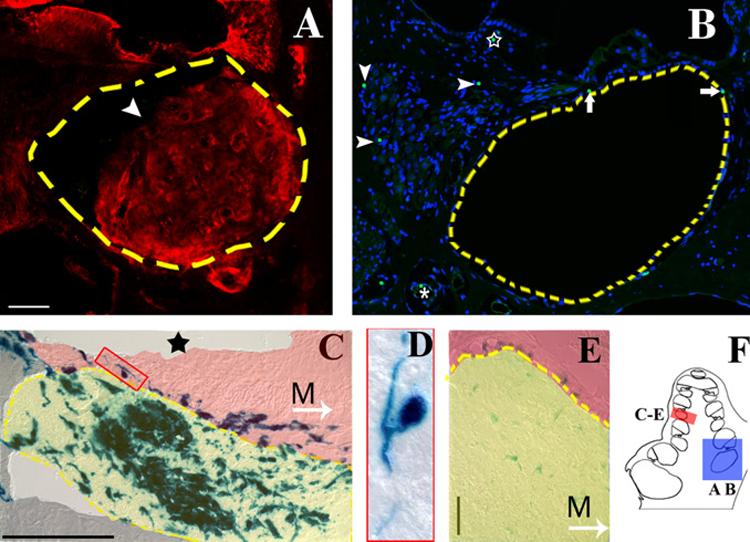
A) Round window injections into the murine cochlea resulted in a bolus of cells (arrowhead) located in the basal turns of the scala tympani (broken line). β-galactosidase positive cells (Texas red) were observed in the bolus itself, as well as in the spiral ganglion, spiral limbus, organ of Corti, and spiral ligament. Nonspecific labeling was observed in the spiral limbus and tectorial membrane. B) Perfusion throughout the cochlea and vestibular system failed to produce obstruction the basal turns of the scala tympani; however, transplanted cells were observed within the cochlear tissue. A mid-turn view of a female guinea pig cochlea shows Y- positive cells (green FITC punctate label localized to the DAPI labeled nucleus in blue) lining the scala tympani (arrows) and blood vessel (asterisk), and within the spiral ganglion (arrow heads) and spiral limbus (star). Blue= nuclear DAPI label, green= FITC labeled Y-chromosome. C) X-gal labeling of a murine cochlea two weeks after transplantation shows that transplanted cells within the scala tympani (highlighted in yellow) primarily exhibit an amorphous morphology. Some cells that migrated into the osseous spiral lamina region (highlighted in red) exhibited a pseudounipolar neural morphology (box). The sound stimulus has ablated any remaining cells of the organ of Corti (star indicates the location of the organ of Corti in a control animal). M, Arrow = direction to modiolus. D) Detail of cell outlined in box of (C). E) X-gal labeling of the bolus within the scala tympani (outlined in yellow) 4 weeks after transplantation illustrates a decrease in LacZ expression over time. The spiral ganglion region is highlighted in red. M, Arrow = direction to modiolus. F) Schematic highlights the representative regions for A and B (blue box) and C-E (red box). Broken line = scala tympani boundary. Scale bars=50 μM.