


Fig. 3.
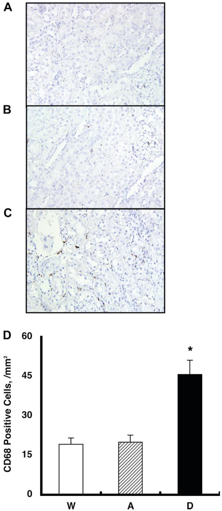
Interstitial macrophage/monocyte infiltration was evaluated by CD68-positive cell number, which is a surface marker for macrophages and monocytes, using zinc-saturated formalin-fixed paraffin-embedded kidney samples from group W (A), group A (B), and group D (C) mice. CD68-positive cells are stained in brown. D: CD68-positive cell numbers were increased significantly in group D mice (46 ± 5 cells/mm2) compared with group A (20 ± 3) and group W (19 ± 2) mice. *P < 0.05 compared with the group W mice.