


Figure 3.
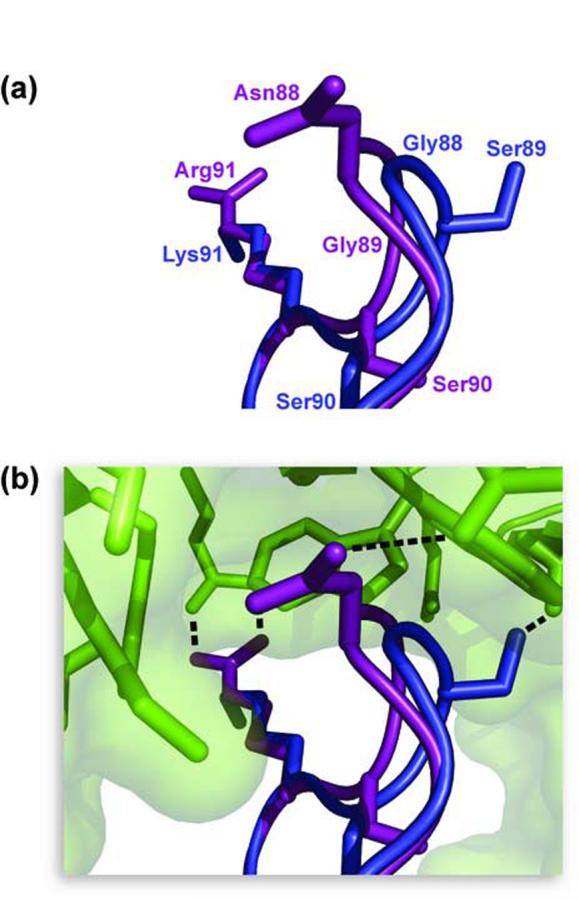
Comparison of the β4-β5 loop of RNase 1 (purple) and RNase A (blue) when bound to RI (green). α-Carbon atoms of RNase 1 and RNase A were superimposed with the program Sequoia, and images were created with the program PyMOL. (a) Structure of the β4-β5 loops. Sidechains of residues 88-91 are shown as sticks. Amino acids are labeled with the color corresponding to the color of the ribonuclease. (b) Orientation of the β4-β5 loops bound to RI. A model of RNase A (PDB entry 1DFJ, chain E) bound to hRI (chain Y) was created based on its alignment to RNase 1 (chain Z). Hydrogen bonds are shown as dotted lines. Hydrogen bonds between hRI and RNase A are hypothesized based on the alignment of hRI (chain Y) and pRI (chain I).