



Abstract
Although the vast majority of genomic DNA is tightly compacted during mitosis, the promoter regions of a number of genes remain in a less compacted state throughout this stage of the cell cycle. The decreased compaction of these promoter regions, which is referred to as gene bookmarking, is thought to be important for the ability of cells to express these genes during the following interphase. Previously, we reported a role for the DNA-binding protein heat shock factor (HSF2) in bookmarking the stress-inducible 70 000-Da heat shock protein (hsp70) gene. In this report, we have extended those studies and found that during mitosis, HSF2 is bound to the HSE promoter elements of other heat shock genes, including hsp90 and hsp27, as well as the proto-oncogene c-fos. The presence of HSF2 is important for expression of these genes because blocking HSF2 levels by RNA interference techniques leads to decreased levels of these proteins. These results suggest that HSF2 is important for constitutive as well as stress-inducible expression of HSE-containing genes.
INTRODUCTION
During mitosis, transcription ceases, most sequence-specific DNA-binding proteins are displaced, and chromatin undergoes condensation (Martinez-Balbas et al 1995; Segil et al 1996). However, the promoters of a number of genes, including those of the inducible form of the 70 000-Da heat shock protein (hsp70i) and c-myc genes, are not tightly compacted in mitotic cells and remain accessible (Martinez-Balbas et al 1995; Michelotti et al 1997; John and Workman 1998; Christova and Oelgeschlager 2002). This lack of compaction of promoter regions in mitotic cells is referred to as “bookmarking” because it is believed to function to permit genes that existed in a transcription-competent state before entry into mitosis to be maintained in a form that can be rapidly reassembled into the active state in G1, akin to the way a bookmark holds a page in a book. A previous study from our laboratory showed that the DNA-binding protein heat shock factor 2 (HSF2) mediates bookmarking of the stress-inducible hsp70 gene by binding to heat shock elements (HSEs) in its promoter in mitotic cells and recruiting the phosphatase PP2A to dephosphorylate nearby condensin complexes, thereby preventing compaction of this region of chromosomal DNA (Xing et al 2005). This work also indicated that sumoylation of the HSF2 protein is higher in mitotic cells compared with asynchronous cells and is important for the ability of HSF2 to interact with the CAP-G subunit of condensin, suggesting that this modification could be a regulator of the HSF2 bookmarking function. The results also showed that HSF2-mediated bookmarking is important for the ability of G1 cells to induce Hsp70 expression in response to stress (Xing et al 2005).
Hsp70 is the best characterized stress-responsive gene with enhanced expression because of 2 HSEs, with most stress-induced expression attributed to the more proximal HSE (Wu et al 1986; Abravaya et al 1991). However, as illustrated in Figure 1, the promoters of a number of other genes also contain HSEs, including those of the important cellular proteins Hsp27, Hsp90, and c-Fos (Hickey et al 1986; Ishikawa et al 1999; Zhang et al 1999). All 3 proteins are responsive to various stresses and protect cells during times of cellular challenge (Wisdom 1999; Jochum et al 2001). However, Hsp90, Hsp27, and c-Fos are also constitutively expressed and have very important functions in cells in the absence of stress (Concannon et al 2003).
Fig 1.

Schematic diagram of the proximal promoters of HSE-containing genes. The proximal promoter regions of the hsp27, hsp70, hsp90, and c-fos genes are shown with the position of the HSE promoter elements labeled relative to the transcription initiation start site (+1). Arrows represent the binding sites for primers used to analyze DNA obtained with chromatin immunoprecipitation assays
For example, the HSP90 family of ATP-dependent chaperones is highly abundant, comprising 1–2% of the protein content in the cytoplasm, and is crucial for the viability of eukaryotic cells (reviewed in Csermely et al 1998). Hsp90 interacts with many different proteins involved in signaling pathways, including steroid receptors, wherein it functions in receptor regulation and likely receptor turnover and trafficking (Pratt and Toft 1997). Hsp70 is important for the ability of Hsp90 to bind to the hormone binding domain of the glucocorticoid receptor (Hutchison et al 1994). Hsp90 is also important for the correct folding and activity of several protein kinases (Csermely et al 1998). Indeed, reduction in levels of Hsp90 leads to low levels of cyclin-dependent kinase Cdc2 and pololike kinase 1 (Plk1) as well as cell cycle arrest mainly at G2 phase following thermal stress (Zou et al 1998; de Carcer 2004). The binding of Hsp90 to HSF1 is thought to keep this stress-responsive transcription factor in a repressed and transcriptionally inactive form (Nakai and Ishikawa 2001).
The molecular chaperone Hsp27 interacts with numerous proteins and is involved in regulation of apoptosis in addition to a number of other important cellular processes (reviewed in Ciocca et al 1993). Mutations in Hsp27 have been linked to various diseases, including Charcot– Marie–Tooth disease and distal hereditary motor neuropathy (Evgrafov et al 2004). High levels of Hsp27 are found in estrogen target organs of the reproductive tract (Ciocca et al 1982, 1983). Overexpression of hsp27 has been reported recently in many kinds of tumorigenic tissues and found to be associated with poor prognosis of astrocytic brain tumor, breast cancer, malignant fibrous histiocytoma, ovarian carcinoma, osteosarcoma, and hepatocellular carcinoma, although the reason for such high levels is not entirely understood (Ciocca et al 1983; Ciocca and Calderwood 2005). Hsp27 has been linked to chemotherapeutic drug resistance (Huot et al 1991) and also appears to function to inhibit apoptosis in cancer cells (Garrido et al 2003; Heijst et al 2005).
The c-fos gene is a member of the FOS family of transcription factors that heterodimerizes with Jun proteins to form the AP-1 (activating protein 1) transcription factor complex. c-Fos is often overexpressed in tumor cells and has oncogenic activity (Schutte et al 1989). Expression of the c-fos gene is enhanced in response to heat, arsenite, and cadmium (Ishikawa et al 1999). AP-1 is involved in regulating genes that are important for a variety of processes, including cell proliferation (Shaulian and Karin 2002). Constitutive activation of AP-1 complex through unregulated c-Fos or c-Jun expression can lead to abnormal cell proliferation and cell transformation (Karin 1995; Karin et al 1997).
In light of the crucial biological functions of Hsp27, Hsp90, and c-Fos, it is important to understand the mechanisms that control the expression of these genes during mitosis. The presence of HSEs in the promoters of all 3 of these genes prompted the question of whether they, like the hsp70i promoter (Xing et al 2005), are bound by HSF2 during mitosis and, if so, whether this binding is important for their expression. The results of this study reveal that HSF2 does bind to the promoters of these genes and is important for the normal level of constitutive expression of these genes. These findings suggest that HSF2 functions as a bookmarking factor for multiple HSE-containing promoters and that it is important not only for stress-induced expression of the hsp70i gene (Xing et al 2005) but also for constitutive expression of the HSE-containing promoters of the hsp90, hsp27, and c-fos genes.
MATERIALS AND METHODS
Materials
Primers were purchased from Integrated DNA Technologies (Coralville, IA, USA). The HSF2 polyclonal antibodies have been described previously (Sarge et al 1993). The secondary antibody, α-IgG-HRP (α-immunoglobulin– horseradish peroxidase), was obtained from an enhanced chemiluminescence (ECL) kit (Amersham Life Science Inc, Arlington Heights, IL, USA). Hsp90 and Hsp27 polyclonal antibodies were purchased from StressGen (Ann Arbor, MI, USA), and the c-Fos antibodies were obtained from Upstate (Charlottesville, VA, USA).
Cell culture
Jurkat cells were grown in RPMI supplemented with 10% fetal calf serum (FCS) and 50 μg/mL gentamicin in a spinner flask at 37°C under 5% CO2. The HeLa cell line (American Type Culture Collection [ATCC, Manassas, VA, USA]) was cultured at 37°C with 5% CO2 in Dulbecco's modified Eagle's medium (DMEM) supplemented with 10% FCS, 2 mM l-glutamine, 4.5 g/L glucose, and 50 μg/mL gentamicin. Mitotic populations of Jurkat cells were obtained by treating cells with Nocodazole at a final concentration of 200 ng/mL for 16 hours, which yields mitotic indices of at least 90%.
Chromatin immunoprecipitations
Chromatin immunoprecipitations (ChIPs) were performed according to established protocols in our laboratory (Xing et al 2005) with the following exceptions. Precleared chromatin was incubated with 5 μL rabbit polyclonal HSF2 antisera or 5 μg control rabbit IgG and rotated at 4°C for 16 hours. DNA was purified with the use of a QIAquick PCR Purification Kit (Qiagen Inc, Valencia, CA, USA) and eluted in 50 μL of H2O (pH 7.5). Aliquots of 2 μL immunoprecipitated DNAs, as well as input samples obtained before immunoprecipitation, were analyzed by polymerase chain reaction (PCR). Amplification conditions were optimized for each set of oligonucleotides. The following primer pairs were used for PCR.
hsp70i
(s) 5′-CAACACCCTTCCCACCGCCACTC-3′
(as) 5′-TGCAGCCGCACAGGTTCGCTCT-3′
hsp27
(s) 5′-CAACCTGTCTGGCTCTGTCC-3′
(as) 5′-GGCAATGACCCGTTTGAGG-3′
hsp90
(s) 5′-GGCGATTGAGGGAAGGTTG-3′
(as) 5′-GGCTAAGTGACCGCACAGGA-3′
c-fos
(s) 5′-GCAGCCCGCGAGCAGTT-3′
(as) 5′-GCCTTGGCGCGTGTCCTAATC-3′
Histone H4
(s) 5′-GAGAGGGCGGGGACAATTGA-3′
(as) 5′-TTGGCGTGCTCGGTGTAGGT-3′
Dihydrofolate reductase
(s) 5′-GGCCTCGCCTGCACAAATAGGG-3′
(as) 5′-GGGCAGAAATCAGCAACTGGGC-3′
Histone H2B
(s) 5′-GCGCGAAAAAGAAGACGTGTTGTT-3′
(as) 5′-TGCGCTCTTGCCGTCTTTCTTCT-3′
β-Globin
(s) 5′-TACCCTAGGGTTGGCCAATCTACT-3′
(as) 5′-CAGCCTAAGGGTGGGAAAATAGAC-3′
Small interfering RNA
HSF2 small interfering RNA (siRNA) was made as previously described (Xing et al 2005). HeLa cells (ATCC) were transfected with 2.5 μg of HSF2 siRNA for 48 hours by Genesilencer transfection reagent (Gene Therapy Systems, San Diego, CA, USA). Cells extracts were prepared with Buffer C (20 mM N-2-hydroxyethylpiperazine-N′-2-ethane-sulfonic acid, pH 7.9, 25% glycerol, 420 mM NaCl, 1.5 mM MgCl2, 0.2 mM ethylenediaminetetraacetic acid, 0.5 mM phenylmethanesulphonylfluoride, 0.5 mM dithiothreitol). Five micrograms of extract was resolved on a 10% sodium dodecyl sulfate–polyacrylamide gel electrophoresis (SDS-PAGE) gel and transferred to nitrocellulose according to standard protocol. The membrane was blocked with 5% nonfat dry milk (NFDM), blotted for HSF2 (1: 5000), HSF1 (1:5000), Hsp27 (1:5000), Hsp90 (1:5000), c-Fos (1:100), and actin (1:10 000) diluted in NFDM. Membranes were incubated with SuperSignal West Pico or Femto ECL reagent (Pierce Biotechnology Inc, Rockford, IL, USA), and bands were visualized by autoradiography.
RESULTS
HSF2 binds hsp27 and hsp90 gene promoters during mitosis
Previous work from our laboratory showed that the DNA-binding protein HSF2 binds to HSEs in the promoter of the hsp70i gene during mitosis and plays a key role in preventing compaction of this region during this stage of the cell cycle, a phenomenon referred to as bookmarking (Xing et al 2005). However, the promoters of other genes, some of which are members of the heat shock protein gene family, including the hsp90 and hsp27 genes, also contain HSEs. The locations of the HSEs in these promoters are shown in Figure 1. This prompted the questions of whether HSF2 might also be binding to the promoters of these other genes during mitosis and, if so, whether this HSF2 function also plays some role in modulating expression of these genes as it does for the hsp70i gene.
To address the first question, we performed ChIP analysis on asynchronous and mitotic (nocodazole-blocked) populations of Jurkat cells with the use of either HSF2 antibodies or species-matched nonspecific IgG (as negative control), followed by PCR analysis of the immunoprecipitated DNA fragments with primers that amplify the promoter regions of the hsp90 and hsp27 genes. Analysis of HSF2 binding to the hsp70i gene promoter was included in this experiment as a positive control. The results of this experiment, shown in Figure 2A, indicate that HSF2 does bind to the promoter regions of the hsp27 and hsp90 genes, with consistently more binding observed in mitotic cells than asynchronous cells. The weaker intensity of the binding signals for the asynchronous cell sample presumably reflects the population of mitotic cells within the asynchronous cell sample. The results in Figure 2A also show that HSF2 does not appear to bind to the promoters of a number of genes that lack HSE promoter elements, including the histone H2B and dihydrofolate reductase genes (H2B and DHFR, respectively), indicating the specificity of the interactions between HSF2 and the hsp90 and hsp27 promoter regions. The specificity of the HSF2 antibodies used in this assay is supported by the lack of PCR product observed when rabbit IgG was used to immunoprecipitate the cross-linked complexes. The input lanes show similar loading and PCR amplification for the different samples.
Fig 2.

Increased mitotic binding of HSF2 but not RNA polymerase II to HSE promoters. (A) Chromatin immunoprecipitation (ChIP) assays were performed with either HSF2 antibodies or nonspecific antibodies (as negative controls) and analyzed by polymerase chain reaction (PCR) with primers that amplify promoter regions of the hsp70, hsp27, hsp90, H2B, and DHFR genes. The top panel for each gene represents a PCR reaction with the use of HSF2-precipitated DNA fragments obtained from an asynchronous or mitotic (nocodazole treated) population of Jurkat cells. The middle panels represent PCR reactions with nonspecific rabbit immunoglobulin (IgG)-precipitated DNA fragments as template from asynchronous and mitotic populations of Jurkat cells. The lower panels represent PCR reactions with template DNA that was treated in the same manner as the HSF2- and IgG-precipitated DNA, except the input DNA was not subjected to immunoprecipitation. (B) ChIP assays with RNA polymerase II antibodies were performed as above and analyzed by PCR with primers that amplify promoter regions of the hsp70, hsp90, and c-fos genes
To further support our finding that HSF2 binding increases in mitotic cells, the binding of RNA polymerase II (Pol II) to the promoters of hsp70, hsp90, and c-fos was analyzed via ChIP assays in asynchronous and mitotic cells. Most factors are displaced from DNA as cells proceed into mitosis, and consistent with this, we found that the binding of RNA Pol II to the promoters of hsp70, hsp90, and c-fos is decreased in mitotic cells (Fig 2B). Interestingly, we found that RNA polymerase II bound to the hsp27 promoter consistently between mitotic and asynchronous populations of cells (data not shown).
hsp27 and hsp90 expression is decreased by HSF2 RNA interference
On the basis of our findings shown above that HSF2 binds to the promoters of the hsp27 and hsp90 genes during mitosis, and our previous finding that HSF2 is important for the ability of cells to express the hsp70i gene (Xing et al 2005), we hypothesized that HSF2 could also be important for expression of these genes. To test this, we reduced cellular HSF2 protein levels by treating an asynchronous population of HeLa cells with HSF2-specific siRNA or scrambled siRNA and determined the effect of this treatment on Hsp27 and Hsp90 protein levels by Western blot analysis (Fig 3). We first assayed the effect of treating HeLa cells with HSF2-specific siRNA or scrambled siRNA on HSF2 and HSF1 levels to confirm that HSF2 levels were specifically reduced (Fig 3A). The results showed that HSF2 was significantly reduced in HSF2 siRNA-transfected cells, but not in cells transfected with the negative control with scrambled siRNA. The protein level of HSF1 was not affected by HSF2-specific siRNA treatment, indicating the specificity of the HSF2 siRNA. We next analyzed the levels of the Hsp27 and Hsp90 proteins in the HSF2-siRNA– and scrambled siRNA–treated HeLa cells and found that the levels of both of these proteins was reproducibly decreased while the level of the control protein β-actin remained constant. The results of this experiment suggest that HSF2 is important for normal levels of constitutive expression of the hsp90 and hsp27 genes.
Fig 3.
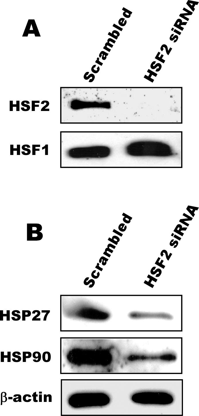
Inhibiting HSF2 expression decreases expression of hsp90 and hsp27. Asynchronous populations of HeLa cells were transfected with HSF2-specific or scrambled small interfering RNA (siRNA), and then extracts of these cells were subjected to sodium dodecyl sulfate–polyacrylamide gel electrophoresis and Western blot analysis with antibodies against HSF2 and HSF1 (A) or against Hsp27, Hsp90, or β-actin (B)
In vivo interactions between HSF2 and c-fos
A number of genes that are not members of the Hsp gene family also have functional HSEs within their promoters. One of these genes is the important proto-oncogene c-fos. Proper regulation of expression of the c-fos gene is crucial for a number of important cellular processes including proliferation; thus, the presence of an HSE in the promoter of this gene prompted us to test whether HSF2 binds the c-fos promoter region in mitotic cells and, if so, whether HSF2 is important for expression of this gene.
As shown in Figure 4A, the results of ChIP assay experiments indicate that HSF2 does interact with the c-fos promoter and that, similar to the results for the hsp90 and hsp27 promoters (Fig 2), or previously for the hsp70i promoter (Xing et al 2005), more HSF2 binding to the c-fos promoter was observed in mitotic cells than asynchronous cells. As in Figure 2, rabbit IgG was used as a control to show the specificity and analysis of input DNAs to control for loading and PCR amplification. The non-HSE–containing histone H4 promoter (H4) was used as a negative control in this experiment.
Fig 4.

HSF2 binds the HSE-containing promoter of the c-fos gene in mitotic cells and is important for expression of this gene. (A) The results of polymerase chain reaction (PCR) with chromatin immunoprecipitation (ChIP) DNA immunoprecipitated with c-Fos and H4 antibodies. The panels for each gene represent PCR reactions carried out as labeled in Figure 2, with the top panel representing HSF2-precipitated DNA fragments used as PCR template, the middle panels representing rabbit immunoglobulin (IgG)-precipitated DNA fragments as template, and the lower panels representing mock-treated template DNA. (B) An asynchronous population of HeLa cells were treated with HSF2-specific or scrambled small interfering RNA (siRNA), and the effect on c-Fos protein levels was assayed by Western blots with c-Fos antibodies. A Western blot with antibodies against β-actin was performed as a loading control
In light of our results showing the importance of HSF2 for expression of the hsp70i gene (Xing et al 2005) and the hsp90 and hsp27 genes (Fig 3), this result showing binding of HSF2 to the c-fos promoter in mitotic cells suggested that HSF2 might also be important for expression of the c-Fos protein. To test this, extracts of asynchronous HeLa cells transfected with the HSF2 siRNA or scrambled siRNA were subjected to Western blot with the use of c-Fos antibodies (Fig 4B). c-Fos can exist in both phosphorylated and unphosphorylated forms, which previous studies indicated are detected on SDS-PAGE as multiple bands between 47 and 70 kDa (Tratner et al 1990; Kovary and Bravo 1991). We found that the HSF2 siRNA treatment reproducibly reduced the levels of c-Fos proteins compared with that observed for extracts of cells treated with the scrambled siRNA negative control. These results suggest that HSF2 is important for normal levels of c-Fos expression in cells.
DISCUSSION
Our previous data showed that HSF2 binds to the promoter of the hsp70i gene in mitotic cells and that HSF2 function is important for the ability of cells in G1 to express the hsp70i gene in response to stress (Xing et al 2005). The results presented in this paper now provide 2 new insights into the activity and functions of HSF2.
First, these results reveal that the binding of HSF2 to HSE promoter elements during mitosis is not exclusive to those in the hsp70i promoter. Our results indicate that during mitosis, HSF2 binds to other promoters that contain HSEs, including the promoters of such genes as hsp90, hsp27, and c-fos. In future studies this list could continue to grow because a number of other promoters have been shown to be bound by HSF2 (Trinklein et al 2004). We also show that the increased binding of HSF2 during mitosis is specific because RNA Pol II is displaced from the promoters of hsp70, hsp90, and c-fos in mitotic cells.
Second, the results described in this paper also suggest that HSF2 binding to HSE-containing promoters is not only important for stress-inducible expression of genes, as was found to be the case for the hsp70i gene, but is also important for normal levels of constitutive expression of HSE-containing genes as well, in this case the hsp90, hsp27, and c-fos genes. This finding is consistent with the hypothesis that the function of HSF2-mediated bookmarking is to help maintain the promoters it binds during mitosis in a state that can be readily reassembled into transcription complexes in G1 so that transcription of these genes can begin again in G1 as soon as possible (Sarge and Park-Sarge 2005). If HSF2 is not present, this would result in a delay in the beginning of expression of these genes until they can become decompacted, resulting in lower total levels of these gene products, which is what we observe. We cannot completely rule out other possible contributions of other HSF2 functions to the expression of these genes or that HSF2 might contribute to some extent at other parts of the cell cycle, but we think our interpretation above is the one best supported by our data.
As detailed above in the introduction, hsp90, hsp27, and c-fos are all important for the ability of cells to proliferate. Thus, the results indicating the importance of HSF2 for normal levels of expression of these 3 genes could provide a potential explanation, at least in part, for the previously observed relationship between HSF2 and cell proliferation. This previous study found that embryonic fibroblasts from HSF2−/− mice proliferate significantly more slowly than embryonic fibroblast cells from HSF2+/+ mice, but the mechanism by which the loss of HSF2 gave rise to these effects was not clear (Paslaru et al 2003). The decreased levels of Hsp90, Hsp27, c-Fos, or a combination of these proteins could also contribute to the elevated stress-induced killing previously observed in cells in which HSF2 levels had been decreased by RNA interference (RNAi) (Xing et al 2005).
We are aware that previous analyses of the HSF2−/− mouse embryonic fibroblasts (MEFs) did not find a significant reduction in Hsp27 in these cells relative to HSF2+/+ MEFs (Paslaru et al 2003), which we corroborated in experiments in our own laboratory with cells kindly provided by Valerie Mezger (data not shown). However, it is important to note that the HSF2−/− MEF cells have been found to exhibit a number of characteristics of stressed cells, including nuclear localization of Hsp70 and the stress-responsive transcription factor HSF1, as well as a significantly lowered temperature threshold for induction of the stress response in these cells compared with HSF2+/+ MEF cells (Paslaru et al 2003). Thus, one explanation for the difference is that the stimulation of stress pathways in the HSF2−/− cells that impinge on the hsp27 and hsp90 promoters could have led to up-regulation of these genes and thereby compensated for potential effects of HSF2 loss on their expression level. Consistent with this possibility, this previous study found that levels of Hsp25 (mouse homolog of human Hsp27) were even higher in HSF2−/− than HSF2+/+ MEF cells. Another possible explanation for the difference between the results of Hsp90 and Hsp27 level results in HSF2−/− MEFs vs HSF2 RNAi-treated cells could be that the MEF cells had adapted to the loss of HSF2 and altered other regulatory pathways to compensate for its loss. Blocking HSF2 by siRNA techniques might provide a more acute analysis of events because the cells presumably do not have as much time to compensate for the loss of HSF2. We also note that, consistent with our results with HSF2 RNAi-treated cells, a previous study did show that c-Fos expression is decreased 4.5-fold in testis cells from HSF2−/− mice compared with HSF2+/+ mice (Wang et al 2004), which alone could provide a potential explanation for the proliferation defect observed in MEF cells.
The results of this paper suggest that HSF2 can bind to the promoters of hsp27, hsp90, and c-fos during mitosis and is important for the normal expression of these genes. Future studies could reveal a role for HSF2 in binding and regulating the expression of additional genes that participate in a variety of downstream cellular processes.
Acknowledgments
We thank the other members of our laboratory for insightful discussions during the course of this work. This work was supported by a Lalor Foundation postdoctoral grant and a National Institutes of Health (NIH) HD grant (F32HD050043) to D.C.W, NIH Training Grant support (ES007266) to H.S.S., and NIH grants GM61053 and GM64606 to K.D.S.
REFERENCES
- Abravaya K, Phillips B, Morimoto RI. Heat shock-induced interactions of heat shock transcription factor and the human hsp70 promoter examined by in vivo footprinting. Mol Cell Biol. 1991;11:586–592. doi: 10.1128/mcb.11.1.586-592.1991.1098-5549(1991)011[0586:HSIOHS]2.0.CO;2 [DOI] [PMC free article] [PubMed] [Google Scholar]
- Christova R, Oelgeschlager T. Association of human TFIID– promoter complexes with silenced mitotic chromatin in vivo. Nat Cell Biol. 2002;4:79–82. doi: 10.1038/ncb733.1465-7392(2002)004[0079:AOHTPC]2.0.CO;2 [DOI] [PubMed] [Google Scholar]
- Ciocca DR, Adams DJ, Bjercke RJ, Edwards DP, McGuire WL. Immunohistochemical detection of an estrogen-regulated protein by monoclonal antibodies. Cancer Res. 1982;42:4256–4258.0008-5472(1982)042[4256:IDOAEP]2.0.CO;2 [PubMed] [Google Scholar]
- Ciocca DR, Adams DJ, Edwards DP, Bjercke RJ, McGuire WL. Distribution of an estrogen-induced protein with a molecular weight of 24,000 in normal and malignant human tissues and cells. Cancer Res. 1983;43:1204–1210.0008-5472(1983)043[1204:DOAEPW]2.0.CO;2 [PubMed] [Google Scholar]
- Ciocca DR, Calderwood SK. Heat shock proteins in cancer: diagnostic, prognostic, predictive, and treatment implications. Cell Stress Chaperones. 2005;10:86–103. doi: 10.1379/CSC-99r.1.1466-1268(2005)010[0086:HSPICD]2.0.CO;2 [DOI] [PMC free article] [PubMed] [Google Scholar]
- Ciocca DR, Oesterreich S, Chamness GC, McGuire WL, Fuqua SA. Biological and clinical implications of heat shock protein 27,000 (Hsp27): a review. J Natl Cancer Inst. 1993;85:1558–1570. doi: 10.1093/jnci/85.19.1558.0027-8874(1993)085[1558:BACIOH]2.0.CO;2 [DOI] [PubMed] [Google Scholar]
- Concannon CG, Gorman AM, Samali A. On the role of Hsp27 in regulating apoptosis. Apoptosis. 2003;8:61–70. doi: 10.1023/a:1021601103096.1360-8185(2003)008[0061:OTROHI]2.0.CO;2 [DOI] [PubMed] [Google Scholar]
- Csermely P, Schnaider T, Soti C, Prohaszka Z, Nardai G. The 90-kDa molecular chaperone family: structure, function, and clinical applications. A comprehensive review. Pharmacol Ther. 1998;79:129–168. doi: 10.1016/s0163-7258(98)00013-8.0163-7258(1998)079[0129:TKMCFS]2.0.CO;2 [DOI] [PubMed] [Google Scholar]
- de Carcer G. Heat shock protein 90 regulates the metaphase– anaphase transition in a polo-like kinase-dependent manner. Cancer Res. 2004;64:5106–5612. doi: 10.1158/0008-5472.CAN-03-2214.0008-5472(2004)064[5106:HSPRTM]2.0.CO;2 [DOI] [PubMed] [Google Scholar]
- Evgrafov OV, Mersiyanova I, and Irobi J. et al. 2004 Mutant small heat-shock protein 27 causes axonal Charcot–Marie–Tooth disease and distal hereditary motor neuropathy. Nat Genet. 36:602–606. [DOI] [PubMed] [Google Scholar]
- Garrido C, Schmitt E, Cande C, Vahsen N, Parcellier A, Kroemer G. HSP27 and HSP70: potentially oncogenic apoptosis inhibitors. Cell Cycle. 2003;2:579–584.1551-4005(2003)002[0579:HAHPOA]2.0.CO;2 [PubMed] [Google Scholar]
- Heijst JW, Niessen HW, Musters RJ, Hinsbergh VW, Hoekman K, Schalkwijk CG. Argpyrimidine-modified heat shock protein 27 in human non-small cell lung cancer: a possible mechanism for evasion of apoptosis. Cancer Lett. 2005;241:309–319. doi: 10.1016/j.canlet.2005.10.042.0304-3835(2005)241[0309:AHSPIH]2.0.CO;2 [DOI] [PubMed] [Google Scholar]
- Hickey E, Brandon SE, Potter R, Stein G, Stein J, Weber LA. Sequence and organization of genes encoding the human 27 kDa heat shock protein. Nucleic Acids Res. 1986;14:4127–4145. doi: 10.1093/nar/14.10.4127.0305-1048(1986)014[4127:SAOOGE]2.0.CO;2 [DOI] [PMC free article] [PubMed] [Google Scholar]
- Huot J, Roy G, Lambert H, Chretien P, Landry J. Increased survival after treatments with anticancer agents of Chinese hamster cells expressing the human Mr 27,000 heat shock protein. Cancer Res. 1991;51:5245–5252.0008-5472(1991)051[5245:ISATWA]2.0.CO;2 [PubMed] [Google Scholar]
- Hutchison KA, Dittmar KD, Czar MJ, Pratt WB. Proof that Hsp70 is required for assembly of the glucocorticoid receptor into a heterocomplex with Hsp90. J Biol Chem. 1994;269:5043–5049.0021-9258(1994)269[5043:PTHIRF]2.0.CO;2 [PubMed] [Google Scholar]
- Ishikawa T, Igarashi T, Hata K, Fujita T. c-fos induction by heat, arsenite, and cadmium is mediated by a heat shock element in its promoter. Biochem Biophys Res Commun. 1999;254:566–571. doi: 10.1006/bbrc.1998.9979.0006-291X(1999)254[0566:CIBHAA]2.0.CO;2 [DOI] [PubMed] [Google Scholar]
- Jochum W, Passegue E, Wagner EF. AP-1 in mouse development and tumorigenesis. Oncogene. 2001;20:2401–2412. doi: 10.1038/sj.onc.1204389.0950-9232(2001)020[2401:AIMDAT]2.0.CO;2 [DOI] [PubMed] [Google Scholar]
- John S, Workman JL. Bookmarking genes for activation in condensed mitotic chromosomes. Bioessays. 1998;20:275–279. doi: 10.1002/(SICI)1521-1878(199804)20:4<275::AID-BIES1>3.0.CO;2-P.0265-9247(1998)020[0275:BGFAIC]2.0.CO;2 [DOI] [PubMed] [Google Scholar]
- Karin M. The regulation of AP-1 activity by mitogen-activated protein kinases. J Biol Chem. 1995;270:16483–16486. doi: 10.1074/jbc.270.28.16483.0021-9258(1995)270[16483:TROAAB]2.0.CO;2 [DOI] [PubMed] [Google Scholar]
- Karin M, Liu Z, Zandi E. AP-1 function and regulation. Curr Opin Cell Biol. 1997;9:240–246. doi: 10.1016/s0955-0674(97)80068-3.0955-0674(1997)009[0240:AFAR]2.0.CO;2 [DOI] [PubMed] [Google Scholar]
- Martinez-Balbas MA, Dey A, Rabindran SK, Ozato K, Wu C. Displacement of sequence-specific transcription factors from mitotic chromatin. Cell. 1995;83:29–38. doi: 10.1016/0092-8674(95)90231-7.0092-8674(1995)083[0029:DOSTFF]2.0.CO;2 [DOI] [PubMed] [Google Scholar]
- Michelotti EF, Sanford S, Levens D. Marking of active genes on mitotic chromosomes. Nature. 1997;388:895–899. doi: 10.1038/42282.1476-4687(1997)388[0895:MOAGOM]2.0.CO;2 [DOI] [PubMed] [Google Scholar]
- Nakai A, Ishikawa T. Cell cycle transition under stress conditions controlled by vertebrate heat shock factors. EMBO J. 2001;20:2885–2895. doi: 10.1093/emboj/20.11.2885.1460-2075(2001)020[2885:CCTUSC]2.0.CO;2 [DOI] [PMC free article] [PubMed] [Google Scholar]
- Paslaru L, Morange M, Mezger V. Phenotypic characterization of mouse embryonic fibroblasts lacking heat shock factor 2. J Cell Mol Med. 2003;7:425–435. doi: 10.1111/j.1582-4934.2003.tb00245.x.1582-1838(2003)007[0425:PCOMEF]2.0.CO;2 [DOI] [PMC free article] [PubMed] [Google Scholar]
- Pratt WB, Toft DO. Steroid receptor interactions with heat shock protein and immunophilin chaperones. Endocr Rev. 1997;18:306–360. doi: 10.1210/edrv.18.3.0303.0163-769X(1997)018[0306:SRIWHS]2.0.CO;2 [DOI] [PubMed] [Google Scholar]
- Sarge KD, Murphy SP, Morimoto RI. Activation of heat shock gene transcription by heat shock factor 1 involves oligomerization, acquisition of DNA-binding activity, and nuclear localization and can occur in the absence of stress. Mol Cell Biol. 1993;13:1392–1407. doi: 10.1128/mcb.13.3.1392-1407.1993.1098-5549(1993)013[1392:AOHSGT]2.0.CO;2 [DOI] [PMC free article] [PubMed] [Google Scholar]
- Sarge KD, Park-Sarge OK. Gene bookmarking: keeping the pages open. Trends Biochem Sci. 2005;30:605–610. doi: 10.1016/j.tibs.2005.09.004.0376-5067(2005)030[0605:GBKTPO]2.0.CO;2 [DOI] [PubMed] [Google Scholar]
- Schutte J, Viallet J, Nau M, Segal S, Fedorko J, Minna J. jun-B inhibits and c-fos stimulates the transforming and trans-activating activities of c-jun. Cell. 1989;59:987–997. doi: 10.1016/0092-8674(89)90755-1.0092-8674(1989)059[0987:JIACST]2.0.CO;2 [DOI] [PubMed] [Google Scholar]
- Segil N, Guermah M, Hoffmann A, Roeder RG, Heintz N. Mitotic regulation of TFIID: inhibition of activator-dependent transcription and changes in subcellular localization. Genes Dev. 1996;10:2389–2400. doi: 10.1101/gad.10.19.2389.0890-9369(1996)010[2389:MROTIO]2.0.CO;2 [DOI] [PubMed] [Google Scholar]
- Shaulian E, Karin M. AP-1 as a regulator of cell life and death. Nat Cell Biol. 2002;4:E131–E136. doi: 10.1038/ncb0502-e131.1465-7392(2002)004[E131:AAAROC]2.0.CO;2 [DOI] [PubMed] [Google Scholar]
- Trinklein ND, Chen WC, Kingston RE, Myers RM. Transcriptional regulation and binding of heat shock factor 1 and heat shock factor 2 to 32 human heat shock genes during thermal stress and differentiation. Cell Stress Chaperones. 2004;9:21–28. doi: 10.1379/481.1.1466-1268(2004)009[0021:TRABOH]2.0.CO;2 [DOI] [PMC free article] [PubMed] [Google Scholar]
- Wang G, Ying Z, and Jin X. et al. 2003 Targeted disruption of the heat shock transcription factor (hsf)-2 gene results in increased embryonic lethality, neuronal defects, and reduced spermatogenesis. Genesis. 36:48–61. [DOI] [PubMed] [Google Scholar]
- Wisdom R. AP-1: one switch for many signals. Exp Cell Res. 1999;253:180–185. doi: 10.1006/excr.1999.4685.0014-4827(1999)253[0180:AOSFMS]2.0.CO;2 [DOI] [PubMed] [Google Scholar]
- Wu BJ, Kingston RE, Morimoto RI. Human HSP70 promoter contains at least two distinct regulatory domains. Proc Natl Acad Sci U S A. 1986;83:629–633. doi: 10.1073/pnas.83.3.629.1091-6490(1986)083[0629:HHPCAL]2.0.CO;2 [DOI] [PMC free article] [PubMed] [Google Scholar]
- Xing H, Wilkerson DC, and Mayhew CN. et al. 2005 Mechanism of hsp70i gene bookmarking. Science. 307:421–423. [DOI] [PubMed] [Google Scholar]
- Zhang SL, Yu J, Cheng XK, Ding L, Heng FY, Wu NH, Shen YF. Regulation of human hsp90 alpha gene expression. FEBS Lett. 1999;444:130–135. doi: 10.1016/s0014-5793(99)00044-7.0014-5793(1999)444[0130:ROHHAG]2.0.CO;2 [DOI] [PubMed] [Google Scholar]
- Zou J, Guo Y, Guettouche T, Smith DF, Voellmy R. Repression of heat shock transcription factor HSF1 activation by HSP90 (HSP90 complex) that forms a stress-sensitive complex with HSF1. Cell. 1998;94:471–480. doi: 10.1016/s0092-8674(00)81588-3.0092-8674(1998)094[0471:ROHSTF]2.0.CO;2 [DOI] [PubMed] [Google Scholar]