


Figure 3.
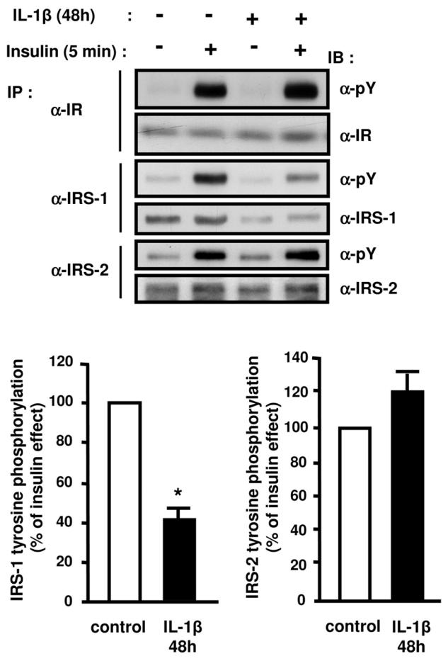
Differential effects of IL-1β treatment on insulin-stimulated tyrosine phosphorylation of the insulin receptor, IRS-1 and IRS-2. 3T3-L1 adipocytes were treated without or with IL-1β (20 ng/ml) for 48 h and then without or with insulin for 5 min. Proteins were immunoprecipitated (IP) using anti-insulin receptor (α-IR) antibodies, anti-IRS1 (α-IRS1) or anti-IRS2 (α-IRS2) antibodies and immunoprecipitated proteins were resolved by SDS-PAGE and blotted (IB) using anti-phosphotyrosine (α-pY) antibodies. The membrane was then stripped and probed using α-IR, α-IRS1, or α-IRS2 antibodies. Typical autoradiographs representative of four to five experiments are shown. The absolute Tyr-phosphorylation of IRS-1 or IRS-2 was quantified by densitometry scanning analysis. Results are expressed as percentage of insulin effect in control cells and presented as the means ± SEM of five independent experiments. *, IL-1β effect significant with p < 0.01.