



Abstract
The hypothalamic paraventricular nucleus (PVN) coordinates major neuroendocrine and behavioral mechanisms, particularly responses to homeostatic challenges. Parvocellular and magnocellular PVN neurons are richly innervated by pituitary adenylate cyclase-activating polypeptide (PACAP) axons. Our recent functional observations have also suggested that PACAP may be an excitatory neuropeptide at the level of the PVN. Nevertheless, the exact localization of PACAP-producing neurons that project to the PVN is not understood. The present study examined the specific contribution of various brain areas sending PACAP innervation to the rat PVN by using iontophoretic microinjections of the retrograde neuroanatomical tracer cholera toxin B subunit (CTb). Retrograde transport was evaluated from hypothalamic and brainstem sections by using multiple labeling immunofluorescence for CTb and PACAP. PACAP-containing cell groups were found to be retrogradely labeled from the PVN in the median preoptic nucleus; preoptic and lateral hypothalamic areas; arcuate, dorsomedial, ventromedial, and supramammillary nuclei; ventrolateral midbrain periaqueductal gray; rostral and midlevel ventrolateral medulla, including the C1 catecholamine cell group; nucleus of the solitary tract; and dorsal motor nucleus of vagus. Minor PACAP projections with scattered double-labeled neurons originated from the parabrachial nucleus, pericoeruleus area, and caudal regions of the nucleus of the solitary tract and ventrolateral medulla. These observations indicate a multisite origin of PACAP innervation to the PVN and provide a strong chemical neuroanatomical foundation for interaction between PACAP and its potential target neurons in the PVN, such as parvocellular CRH neurons, controlling physiologic responses to stressful challenges and other neuroendocrine or preautonomic PVN neurons.
Indexing terms: hypothalamic paraventricular nucleus, PACAP, retrograde tract tracing, stress, parvocellular PVN, magnocellular PVN
The hypothalamic paraventricular nucleus (PVN) is critically important for the regulation of neuroendocrine, autonomic, and behavioral mechanisms, particularly responses to homeostatic challenges (Swanson and Sawchenko, 1980; Sawchenko et al., 1996; Herman et al., 2002). Significant efforts have been focused on characterizing the role of pituitary adenylate cyclase-activating polypeptide (PACAP), among the many peptide transmitters present in this hypothalamic nucleus. Since its isolation from ovine hypothalamic tissue by Akira Arimura’s research team in 1989 (Miyata et al., 1989), PACAP has been found to be widely distributed in the central nervous system (Arimura et al., 1991; Masuo et al., 1993; Arimura, 1998; Hannibal, 2002). Experimental data available so far indicate that intracerebroventricular (icv) or local brain injections of PACAP induce a range of autonomic, behavioral, and neuroendocrine responses, many of which are related to stress and the activation of PVN neurons (Morley et al., 1992; Murase et al., 1993; Chance et al., 1995; Agarwal et al., 2005; Norrholm et al., 2005).
Particularly high densities of PACAP-immunoreactive nerve terminals innervate parvocellular corticotropin-releasing hormone (CRH; Legradi et al., 1998) and thyrotropin-releasing hormone (TRH) neurons (Legradi et al., 1997), and magnocellular vasopressin neurons receive medium-density PACAP input (Shioda et al., 1997), which may underlie some of the responses observed in vivo. A subset of PACAP-containing nerve terminals innervating parvocellular PVN neurons likely originates from medullary catecholamine neurons, insofar as they colocalize with the adrenergic marker phenylethanolamine N-methyltransferase (PNMT; Legradi et al., 1997). Because of the high density, unique distribution, and heterogeneous morphology of PACAP nerve terminals in the PVN (Legradi et al., 1997; Hannibal, 2002), several distinct brain sites should be considered as potential sources of PACAPergic innervation to the PVN. Locations of PACAP-immunoreactive neurons in hypothalamic and extrahypothalamic brain sites include the preoptic area, dorsomedial and arcuate hypothalamic nuclei, pontine parabrachial nucleus, nucleus of the solitary tract, dorsal motor vagal nucleus, ventrolateral medulla, and area postrema (Legradi et al., 1994b; Piggins et al., 1996; Legradi et al., 1997; Hannibal, 2002). Although many of these regions project to the PVN (McKellar and Loewy, 1981; Moga and Saper, 1994), the exact brain site-specific contribution of PACAP-producing neurons to the innervation of the PVN is not known. Therefore, in the present study we identified the specific brain areas sending PACAP innervation to the PVN by using the combination of retrograde neuroanatomical tract tracing and immunohisto-chemistry.
MATERIALS AND METHODS
Animals
Thirty-four adult, male Sprague-Dawley rats (250–380 g; Taconic Farms, Germantown, NY) were used for this study. Animals were housed under controlled environmental conditions (lights on 0600–1800 hours, temperature 22°C, with food and water available ad libitum). All experimental protocols were approved by the Institutional Animal Care Committee of the University of South Florida.
Neuroanatomical retrograde tracing
Rats were anesthetized with a mixture of ketamine (90 mg/kg body weight) and xylazine (10 mg/kg body weight). Rats were mounted in a stereotaxic apparatus (Kopf Instruments) with Bregma and lambda positioned in a horizontal plane. The coordinates used for the PVN were anteroposterior (AP) –1.7 mm from Bregma, lateral 0.4 mm and ventral 8.0 mm from the skull surface. Burr holes were drilled at this position, and glass micropipettes with 30–40-μm tip diameter filled with 0.5% solution of cholera toxin B subunit (CTb; product 104, lot 10426A; List Biologicals, Campbell, CA) in 0.05 M sterile sodium phosphate buffer (PB) were inserted into the brain. CTb was delivered iontophoretically for 6–7 minutes to the PVN by using a positive current of 6 μA with a 7-second on/off pulse. The micropipette was left undisturbed for 10 minutes to avoid diffusion of tracer along the pipette track. Ketoprofen (5 mg/kg) was given to the rats to reduce postsurgical pain and inflammation. After a survival period of 8–10 days, the animals were reanesthetized with ketamine and xylazine and mounted in a stereotaxic apparatus, and 200 μg of colchicine (Sigma, St. Louis, MO) in sterile physiological saline (10 μg/μl) was injected into the lateral ventricle (coordinates: AP – 0.8 mm from Bregma, lateral 1.4 mm ventral 4.6 mm from skull surface) with a stereotaxically positioned Hamilton microsyringe. Ketoprofen (5 mg/kg) was administered to minimize postsurgical pain and inflammation. After a survival of 1.5–2 days, rats were deeply anesthetized with Nembutal (100 mg/kg) and perfused through the ascending aorta with 20 ml heparinized saline followed by 150–200 ml of 4% paraformaldehyde in 0.1 M PB (pH 7.4). Brains were removed, collected in PBS, and cryoprotected overnight in 20% sucrose in PBS at 4°C. Brain blocks were frozen on dry ice and a series of one-in-eight coronal sections at 30-μm thickness were cut with a cryostat to produce sets of sections taken at 240-μm intervals. Sections were collected in PBS, then transferred into freezing solution containing 30% ethylene glycol, 25% glycerol in 0.05M PB at –20°C until further processing.
Evaluation of injection sites
From each brain, one set of sections taken from the hypothalamus was processed for single-labeling immuno-histochemistry for CTb, in some cases followed by neutral red or cresyl violet counterstaining. In an adjacent set of PVN sections, multiple-labeling immunofluorescence was utilized to detect immunoreactivity for CTb and parvo-and magnocellular PVN markers (CRH, vasopressin, or oxytocin).
Single-labeling immunohistochemistry for CTb
Sections were rinsed with PBS and then treated with 0.5% H2O2 in PBS to inhibit endogenous peroxidase activity and to enhance antibody penetration, by 0.5% Triton X-100 in PBS. Nonspecific immunolabeling was reduced by preincubation in 10% normal horse serum (NHS) in PBS. Sections were then incubated with an antiserum raised in goat against CTb (product 703, lot 7032G; List Biologicals) at a dilution of 1:30,000–60,000 in 2% NHS overnight at room temperature, followed by biotinylated donkey anti-goat IgG (at 1:400; Vector Laboratories, Burlingame, CA) for 2 hours at room temperature and then ABC Vector Elite Kit (Vector Laboratories) at 1:100 in PBS for 1 hour. Each incubation step was separated by three 15-minute rinses with PBS. Sections were washed once in 0.05 M Tris buffer (pH 7.6) and then incubated with 0.025% diaminobenzidine (DAB) solution in 0.05 M Tris buffer and 0.0036% H2O2 to visualize CTb immunolabeling similarly to previously published methods (Mihaly et al., 2001). Sections were mounted onto Superfrost Plus slides (Fisher Scientific Co., Pittsburgh, PA), air dried, dehydrated with alcohol followed by xylene, and coverslipped with Cytoseal 60 mounting medium (Richard-Allan Scientific, Kalamazoo, MI). A subset of PVN sections from each brain was also counterstained with neutral red or cresyl violet before coverslipping to aid in delineation of neuroanatomical boundaries.
Double-labeling immunohistochemistry for CTb and PVN markers
Sections were pretreated with 0.5% H2O2 and 0.5% Triton X-100 as described above. Sections were then treated with 10% NHS for 2 hours to reduce nonspecific background and then incubated at 4°C for 3 days with primary antiserum cocktail of goat anti-CTb antiserum (at 1:10,000; List Biologicals) combined with one of the following antisera: guinea pig anti-CRH (at 1:6,000; Peninsula Laboratories, Belmont, CA) rabbit antivasopressin (1:4000; Chemicon, Temecula, CA), rabbit antioxytocin (at 1:4,000; Chemicon). This was followed by an incubation in a cocktail of biotinylated donkey anti-goat IgG (1:400; Jackson Immunoresearch, West Grove, PA) and AMCA-conjugated donkey anti-guinea pig or donkey anti-rabbit IgG (1:50; Jackson Immunoresearch) for 2 hours at room temperature, followed by incubation in Texas red-conjugated avidin (at 1:250; Vector Laboratories) for 2 hours. Each incubation step was separated by three 15-minute washes in PBS. Sections were mounted on glass slides with Vectashield mounting medium (Vector Laboratories) and visualized under an Olympus BX51 fluorescence microscope with specific filters. CTb immunolabeling appeared red, and CRH, vasopressin, or oxytocin immunolabeling appeared blue.
Multiple-labeling immunofluorescence for PACAP, CTb, and PNMT
Series of forebrain and brainstem sections were treated with 0.5% H2O2 and 0.5% Triton X-100 as described above. To obtain optimal PACAP immunolabeling, the sections were heated at 80°C in 10 mM sodium citrate solution, pH 8.5, for 15 minutes to retrieve antigens masked by aldehyde fixation (Jiao et al., 1999). The sections were then treated with 2% nonfat dry milk for 30 minutes and 2% NHS for 1 hour to reduce nonspecific background staining. The sections were then incubated in a primary antiserum cocktail by a well-characterized antiserum made in rabbit against PACAP38 (Piggins et al., 1996; Norrholm et al., 2005; at 1:8,000; catalog T-4473, lot 030354-1; Bachem/Peninsula Laboratories) and goat anti-CTb serum (at 1:10,000) for 3–5 days at 4°C, followed by a second antibody cocktail of biotinylated donkey anti-goat IgG (1:400; Jackson Immunoresearch) and fluorescein isothiocyanate (FITC)-conjugated anti-rabbit antibodies (1: 50; Jackson Immunoresearch) for 4–6 hours at room temperature, followed by Texas red-conjugated avidin (1:250; Vector Laboratories) overnight at 4°C. Sections were mounted and viewed as described above. CTb immunolabeling appeared red, and PACAP immunolabeling appeared green. In control sections, PACAP immunoreactivity was abolished by preadsorption of the primary antiserum with 10 μM concentration of synthetic PACAP (American Peptide Company, Sunnyvale, CA). In a series of sections of the medulla oblongata, triple immunolabeling was used following pretreatment as described above, and the cocktail of PACAP and CTb antisera was also mixed with a guinea pig PNMT antiserum (1:5,000; Research Diagnostics, Flanders, NJ; RDI-PRO16067, lot DS6207d). After incubation in the primary antiserum cocktail, sections were sequentially incubated in a mixture of biotinylated donkey anti-goat IgG (at 1:400), FITC anti-rabbit (at 1:50; Jackson Immunoresearch), and AMCA-conjugated anti-guinea pig IgG (at 1:50; Jackson Immunoresearch) for 4–6 hours at room temperature, followed by Texas red-conjugated avidin (1:250; Vector Laboratories) overnight at 4°C. Sections were mounted onto uncoated glass slides and coverslipped with Vectashield mounting medium or in some cases Vectashield with DAPI (Vector Laboratories) to facilitate identification of underlying nuclear boundaries.
The resultant fluorescence was visualized with the appropriate filter combinations for each marker in an Olympus BX51 microscope, and images were taken with an Olympus Q-Color5 digital microscope camera. After image capture, brightness and contrast were modified and the filter Unsharp mask was applied as necessary, in Adobe Photoshop 7 running on a Power Macintosh G4 computer. Images containing individual fluorescent labeling for each field were superimposed in Photoshop to obtain composite views of single labeled as well as colocalizing neurons. For each anatomically identifiable region, by using cytoarchi-tectonic and immunohistochemical landmarks, numbers of CTb and PACAP-labeled neurons were counted from single sections representing distinct rostrocaudal levels from brains with successful injections. These sections were visually matched to corresponding atlas levels for each brain, with standard rat brain atlases (Swanson, 1998; Paxinos and Watson, 2005), and, for comparisons among different brains, each section was matched with its counterpart within 200 μm or less using atlas AP coordinates. In addition, camera lucida-like drawings were created by importing montages of Photoshop images taken with ×10 (forebrain) or ×4 (brainstem) objectives into the technical illustration program Canvas 8.0 (ACD Systems of America Inc., Miami, FL), where relevant anatomical boundaries and the location of immunolabeled neurons were drawn to visualize the brain ipsilateral to the injection site.
Antiserum characterization and immunolabeling controls
The polyclonal goat anti-CTb (List Biologicals; product 703, lot 7032G) was raised against CTb, a subunit of cholera toxin containing five identical polypeptide chains in a ring-like structure with a total molecular weight of ˜58,000 kDa. On Western blot, this antiserum recognizes the whole CTb molecule when developed with anti-goat antibodies and an appropriate chromogen, according to information provided by List Biologicals. In our laboratory, preadsorption of the CTb antiserum by 10 μM of CTb completely abolished immunolabeling, whereas clear immunolabeling was observed in adjacent sections processed simultaneously with the same antibody concentration without preadsorption.
The anti-PACAP polyclonal serum (Bachem/Peninsula Laboratories; catalog T-4473, lot 030354-1) was made in rabbit against PACAP38 having the amino acid sequence of HSDGIFTDSYSRYRKQMAVKKYLAAVLGKRYKQR-VKNK-NH2. According to Bachem, based on cross-reactivity data obtained by radioimmunoassay, this antiserum binds the C-terminal end of the PACAP38 peptide and does not show any cross-reaction with vasoactive intestinal peptide, CRH, or adrenocorticotrophic hormone and only 0.01% cross-reactivity with PACAP27. In our laboratory, preadsorption of the working dilution of the primary antiserum with 10 μM concentration of synthetic PACAP38 (American Peptide Company; catalog 34-0-20, lot T11107T1), molecular weight 4534.3, completely abolished immunoreactivity. The anti-CRH polyclonal serum (Bachem/Peninsula Laboratories, catalog T-5007, lot GHC8561) was made in guinea pig against a synthetic CRH having an amino acid sequence of SEEPPISLDLTFHLLREVLEMARAEQLAQQAHSNRKL-MEII-NH2. In our laboratory, preadsorption of this antiserum by 10 μM of synthetic CRH (American Peptide Company; catalog 34-1-10, lot T11107T1), molecular weight 3826.4, completely abolished immunolabeling. Antivasopressin (Chemicon; catalog AB1565, lot 25020039) and antioxytocin (Chemicon; catalog AB911, lot 25020806) sera were made in rabbits against full-length vasopressin or oxytocin conjugated to thyroglobulin. Because these hormones are only nine amino acids in length, the binding site of the antibody encompasses the entire molecule. Chemicon tested the specificity of the immunogen conjugates in Western blot for cross-reactivity, using both vasopressin-thyroglobulin and oxytocin-thyroglobulin to verify that the antioxytocin serum does not react with vasopressin and that the antivasopressin serum does not react with oxytocin. In our laboratory, we ran control tests by preadsorbing the antisera using their synthetic peptide antigens at 10 μM, which resulted in a complete loss of immunolabeling. The polyclonal antiphenylethanolamine-N-methyltransferase (PNMT) antiserum (Research Diagnostics; catalog RDI-PRO16067) was raised in guinea pig against PNMT from bovine adrenal medulla and tested by immunolabeling of rat adrenal gland tissue by Research Diagnostics. Preadsorption of the antiserum with 10 or 100 μM PNMT (bovine PNMT; Sigma) in our laboratory completely abolished immunoreactivity as tested on sections of rat medulla oblongata in the present study.
RESULTS
Injection sites
PVN injection sites were initially detected by CTb immunolabeling using the avidin-biotin-immunoperoxidase method visualized with DAB and then further evaluated with immunofluorescence methods. With DAB as chromogen, the core of each injection site appeared as a solid, dense area surrounded by a peripheral zone of diffused tracer. A total of nine cases produced sites in which CTb deposit cores were found fully or partially within the PVN. Among these, five injection sites were disqualified based on the localization and/or the spread of the tracer; either because the tracer deposit core extended significantly beyond the PVN or, conversely, covered only a small portion of the PVN.
In each of the remaining four animals (Nos. 452702, 450702, 511002, and 520301), the injection site was centered at the mid-PVN level (AP: –1.7 to –1.8 mm from Bregma) and covered both the magnocellular and the parvocellular portions of the PVN, with only minimal leakage in the peri-PVN area, reminiscent of a previously published report that also used CTb to delineate major projections to the PVN (Elmquist and Saper, 1996).
A typical injection site is presented in Figure 1A-C(No. 520301), and a composite drawing of all four successful injection sites is shown in Figure 1D. Figure 1A,B illustrates the dense core of the injection at mid- and midcaudal PVN levels, whereas Figure 1C shows the periphery of the injection site with intra-PVN transport in nerve processes of the caudal parvocellular subdivision. The localization of the injection site was evaluated by counterstaining with cresyl violet or neutral red in DAB-labeled sections and by immunofluorescence for CRH or vasopressin or oxytocin cell groups as PVN markers. Fluorescent double immunolabeling for CTb/CRH and CTb/vasopressin or CTb/oxytocin confirmed that CTb injections in these animals covered the medial parvocellular, dorsal, and ventral parvocellular subdivisions and a significant portion of the magnocellular compartment of the PVN, and the injections were confined within the boundaries of the PVN (Fig. 2). Therefore, by using these criteria, the four animals with successful whole PVN injections were considered for analysis of the distribution of PACAP neurons projecting to the PVN.
Fig. 1.
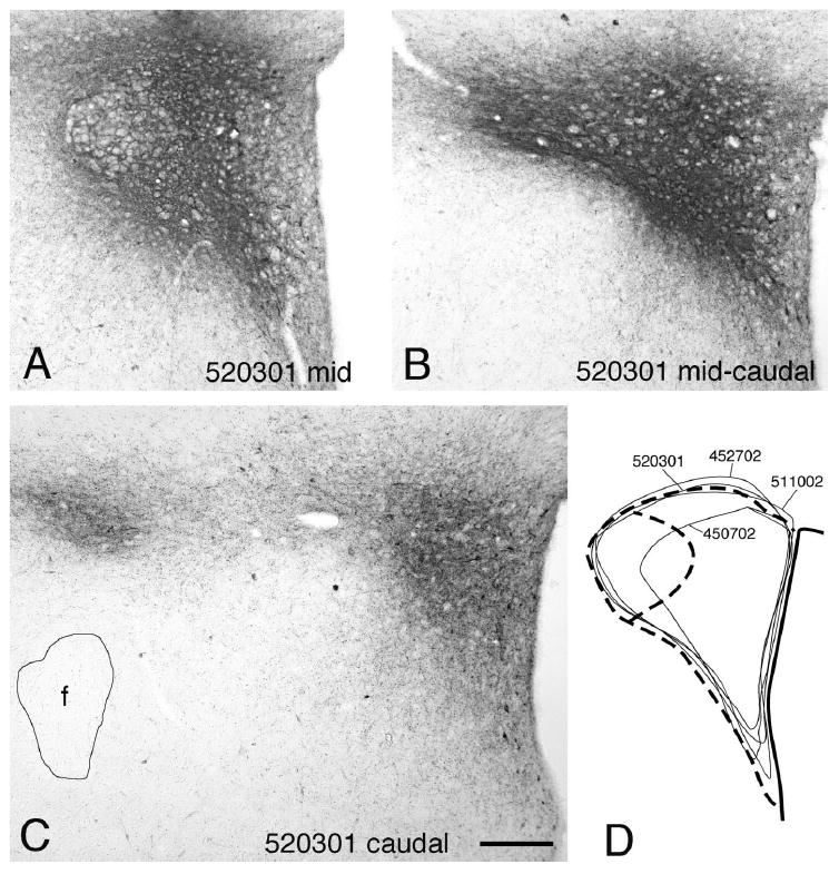
CTb injection sites. A–C: Photomicrographs showing a typical CTb injection site in the PVN (No. 520301) in three coronal levels rostrocaudally, labeled with the immunoperoxidase method with diaminobenzidine as chromogen. D: Summary drawing of successful PVN injection sites centered at midlevel of the PVN (Nos. 450702, 452702, 511002, 520301). Scale bar = 200 μm.
Fig. 2.
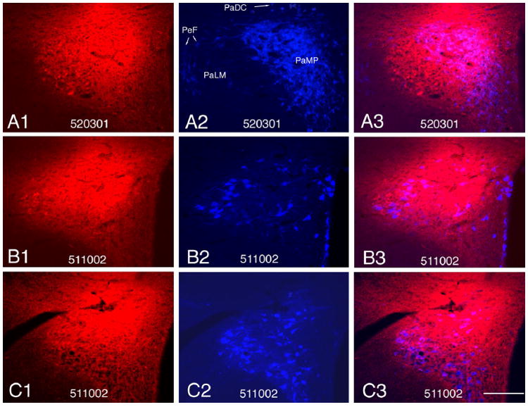
Fluorescent images from two distinct cases (Nos. 511002, 520301) showing the localization of CTb (red fluorescence) injection sites with CRH, oxytocin, or vasopressin (blue fluorescence) neurons as markers of PVN neurons. A1–A3: A CTb injection site using CRH as marker of PVN; A1: CTb injection site; A2: CRH neurons; A3: superimposition of CTb and CRH immunolabeling showing the localization of the injection in the PVN. B1–B3: A CTb injection site using oxytocin as marker of the PVN; B1: CTb injection site; B2: oxytocin neurons in the PVN; B3: superimposition of CTb and oxytocin immunolabeling showing the localization of the injection. C1–C3: A CTb injection site using vasopressin as marker of the PVN; C1: CTb injection site; C2: vasopressin neurons; C3: superimposition of CTb and vasopressin immunolabeling showing the localization of the injection. Scale bar = 200 μm.
Distribution of retrogradely labeled CTb neurons from preoptic level to medulla oblongata
Retrogradely labeled CTb-positive neurons were found in numerous hypothalamic, midbrain, and lower brainstem regions, with strong ipsilateral dominance. Major hypothalamic regions retrogradely labeled from PVN included the parastrial, septohypothalamic, median preoptic (MnPO), medial preoptic (MPO), ventromedial preoptic (VMPO), suprachiasmatic (SCh), retrochiasmatic (RCh), arcuate, dorsomedial (DMN), ventromedial (VMN) nuclei; the medial part of the supramammillary nucleus (SuMM); and medial preoptic (MPA), anterior (AH), perifornical, lateral (LH), and posterior hypothalamic (PHA) areas. Extrahypothalamic forebrain areas containing retrogradely labeled neurons included the anterior-lateral part of the medial subnucleus of the bed nucleus of stria terminalis (STMAL), paraventricular nucleus of the thalamus, and central and medial nuclei of the amygdala. The periventricular gray area (PVG) at the transition between the hypothalamus and the midbrain and the precommissural nucleus also contained retrogradely labeled neurons. Among brainstem regions, retrogradely labeled neurons were observed in the midbrain periaqueductal gray (PAG), lateral parabrachial nucleus and pericoeruleus area, ventrolateral medulla (VLM), nucleus of the solitary tract (NTS), and dorsal motor nucleus of the vagus (DMV). The overall pattern of retrogradely labeled cell bodies was in agreement with previous studies describing PVN-projecting neurons (McKellar and Loewy, 1981; Moga and Saper, 1994; Cullinan et al., 1996; Elmquist and Saper, 1996). Because CTb is also transported anterogradely, CTb-labeled nerve fibers were observed in the external and internal layers of the median eminence and pituitary stalk, indicating their origin from neuroendocrine neurons of the PVN.
General distribution of PACAP neurons
In sections of colchicine-treated brains, at rostrocaudal levels within the scope of the current study, the distributional pattern of PACAP-positive perikarya showed good agreement with previous comprehensive descriptions of PACAP immunolabeling (Legradi et al., 1994a,b; Dun et al., 1996; Piggins et al., 1996; Hannibal, 2002). Briefly, PACAP perikarya were observed in several preoptic nuclei and the medial preoptic area; septohypothalamic nucleus; lateral hypothalamic area; and para- and periventricular, arcuate, dorsomedial, ventromedial, premammilllary, and supramammillary nuclei. In the thalamus, the lateral and medial habenular nuclei contained PACAP-positive cell bodies. Furthermore, PACAP-positive neuronal groups were observed in the cingulate and piriform cortices and at the hypothalamus–midbrain junction; PAG; pontine nuclei; parabrachial nucleus; A5 cell group; locus coeruleus; and dorsal and ventral medulla, including the dorsal motor vagal nucleus (DMV) and nucleus of the solitary tract (NTS); A1/C1, C2, and C3 areas; raphe obscurus and pallidus; ambiguus and lateral reticular nuclei; as well as spinal and medial vestibular nuclei.
Specific localization of PACAP/CTb double-labeled perikarya
Ipsilateral contribution
The specific contribution of PACAP-containing input to the PVN was identified by the presence of simultaneous immunolabeling for PACAP and CTb. PACAP/CTb double-immunolabeled neurons were observed in several hypothalamic regions, including the preoptic and lateral hypothalamic areas; dorsomedial, ventromedial, and arcuate nuclei; ventrolateral column of midbrain PAG; and cell groups in the dorsal and ventro-lateral medulla oblongata. Examples of fluorescent PACAP/CTb immunolabeling in hypothalamic and midbrain regions are shown in Figure 3. At preoptic levels (–0.3 to –0.4 mm from Bregma), the MnPO, MPO, and MPA contained relatively high proportions of PACAP neurons colocalizing with CTb, indicating projections to the PVN (Figs. 3A, 4A). In the bed nucleus of the stria terminalis, significant accumulations of PACAP nerve fibers were found, but CTb and PACAP neurons were observed in separate populations such that PACAP neurons were located in the anteromedial division of the medial subnucleus (STMAM), and CTb neurons were present in the STMAL, but no colocalization was observed. Likewise, in the paraventricular nucleus of the thalamus, CTb-labeled perikarya were readily observed, but only PACAP nerve fibers were present, which is similar to previous findings (Koves et al., 1991; Legradi et al., 1997; Hannibal, 2002). The subfornical organ was not evaluated, because this delicate group of cells was wrinkled and could not be maintained through the rigors of tissue section heating and elongated processing times required for antigen unmasking and double-labeling immunohistochemistry.
Fig. 3.
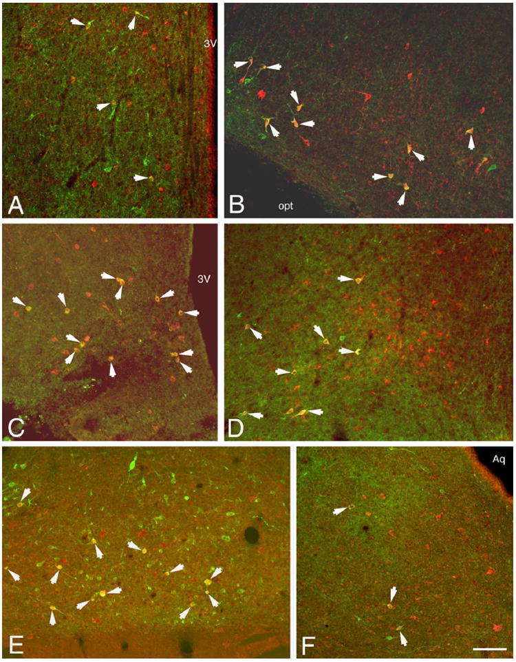
Fluorescent images showing examples of CTb or PACAP or PACAP and CTb double-labeled neurons in selected forebrain and midbrain areas. PACAP neurons show green fluorescence, CTb labeling shows red fluorescence, and double-labeled neurons are indicated by the presence of a composite yellow/orange color (arrows). A: Medial preoptic nucleus. B: Anterior tuberal lateral hypothalamic area. C: Arcuate nucleus. D: Dorsomedial nucleus. E: Supramammillary nucleus. F: Ventrolateral periaqueductal gray. Scale bar = 100 μm.
Fig. 4.
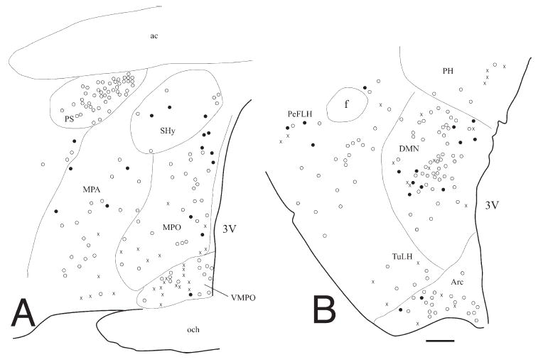
Schematic maps showing the distribution of the Ctb, PACAP, or Ctb-PACAP double-labeled neurons in representative levels of coronal rat hypothalamic sections ipsilateral to the injection site. A: Preoptic region. B: Caudal hypothalamus (–3.5 mm from Bregma). Symbols: ×, PACAP only; open circles, Ctb only; solid circles, PACAP/Ctb double-labeled neurons. Scale bar = 200 μm.
At anterior hypothalamic levels, the suprachiasmatic nucleus, rostral portions of the retrochiasmatic nucleus, and the anterior hypothalamic area lacked PACAP-positive neurons but contained CTb-positive neurons. More caudally, the lateral hypothalamic area, arcuate nucleus, and ventromedial nucleus were among the regions containing significant populations of PVN-projecting PACAP neurons (Fig. 3B,C, Table 1). Within the lateral hypothalamic area (LH), retrogradely labeled PACAP neurons were typically found in a region slightly mediodorsal to the optic tract (Fig. 3B), previously named the intermediate hypothalamic area (Larsen et al., 1994), which corresponds to the tuberal portion of the LH in the standard rat brain atlas (Paxinos and Watson, 2005), and the caudal perifornical lateral hypothalamus (PeLH in Fig. 4B). At tuberal levels of the hypothalamus, the strongest PACAP projections to the PVN were detected at the caudal level of the dorsomedial nucleus (DMN, AP: –3.2 to –3.5 mm from Bregma; Fig. 3D, Table 1), whereas the more rostral portion of the DMN (AP: –2.9 mm) contained a dense network of PACAP-labeled nerve fibers and fewer PACAP/CTb-positive neurons (Table 1).
TABLE 1.
Semiquantitative Evaluation of Retrogradely Labeled PACAP Neurons in Sections Representing Hypothalamic and Brainstem Sources of PACAP Projections to the PVN1
| Number of labeled neurons | |||||
|---|---|---|---|---|---|
| Brain area | CTb | PACAP | Double labeled | Percentage CTb neurons containing PACAP | Percentage PACAP neurons containing CTb |
| Preoptic region | |||||
| Median preoptic nucleus (Bregma –0.4) | 14.3 ± 6.0 | 8.0 ± 2.5 | 4.3 ± 2.8 | 26.4 ± 18.0 | 44.5 ± 17.5 |
| Medial preoptic nucleus (Bregma –0.4) | 36.0 ± 10.4 | 11.7 ± 4.7 | 3.0 ± 1.4 | 7.3 ± 2.7 | 42.9 ± 9.5 |
| Medial preoptic area (Bregma –0.4) | 82.0 ± 57.6 | 20.7 ± 9.2 | 5.5 ± 2.2 | 14.3 ± 6.4 | 42.9 ± 19.0 |
| Ventromedial preoptic nucleus (Bregma –0.4) | 36.6 ± 12.8 | 11.0 ± 3.2 | 2.0 ± 0.6 | 8.3 ± 5.2 | 18.1 ± 0.9 |
| Lateral hypothalamus (Bregma –2.04) | 38.0 ± 9.8 | 21.7 ± 8.4 | 11.3 ± 4.7 | 29.6 ± 8.9 | 59.5 ± 16.2 |
| Ventromedial hypothalamic nucleus (Bregma –2.3) | 28.0 ± 6.6 | 17.6 ± 13.7 | 8.3 ± 5.8 | 24.3 ± 12.8 | 57.0 ± 6.6 |
| Arcuate nucleus | |||||
| Rostral (Bregma –2.3) | 22.7 ± 3.2 | 10.7 ± 2.9 | 2.0 ± 0.6 | 9.2 ± 3.3 | 19.7 ± 5.3 |
| Caudal (Bregma –3.5) | 39.0 ± 9.0 | 10.5 ± 4.5 | 7.5 ± 3.5 | 22.5 ± 14.2 | 70.0 ± 3.3 |
| Dorsomedial hypothalamic nucleus | |||||
| Midlevel (Bregma –2.9) | 48.0 ± 16.0 | 13.0 ± 1.1 | 3.7 ± 1.4 | 8.9 ± 4.9 | 26.6 ± 9.2 |
| Caudal (Bregma –3.4) | 94.6 ± 24.3 | 18.7 ± 3.3 | 11.0 ± 4.6 | 13.5 ± 5.6 | 60.1 ± 19.9 |
| Supramammillary nucleus (Bregma –4.8) | 18.3 ± 5.8 | 14.7 ± 3.9 | 11.3 ± 5.6 | 64.5 ± 24.4 | 28.1 ± 3.4 |
| Ventrolateral periaqueductal gray (Bregma –6.8) | 20.0 ± 4.0 | 9.0 ± 0.2 | 3.5 ± 0.5 | 18.7 ± 6.2 | 38.9 ± 5.6 |
| Lateral parabrachial nucleus (Bregma –9.3) | 24.0 ± 5.5 | 20.3 ± 0.7 | 1.0 ± 0.6 | 4.5 ± 3.1 | 4.9 ± 2.8 |
| Pericoeruleus area (Bregma –9.3) | 14.7 ± 3.4 | 11.3 ± 1.5 | 1.7 ± 0.7 | 11.8 ± 3.6 | 13.9 ± 3.8 |
| Ventrolateral medulla | |||||
| Rostral (Bregma –12.0) | 18.7 ± 6.3 | 18.0 ± 3.1 | 9.7 ± 3.6 | 58.4 ± 10.8 | 52.8 ± 13.5 |
| Midlevel (Bregma –13.3) | 6.2 ± 1.4 | 9.2 ± 2.4 | 4.0 ± 0.6 | 73.3 ± 15.9 | 49.2 ± 9.0 |
| Caudal (Bregma –14.2) | 2.0 ± 1.0 | 3.0 ± 0.6 | 0.6 ± 0.3 | 22.2 ± 11.1 | 19.4 ± 10.0 |
| Nucleus of the solitary tract | |||||
| Rostral (Bregma –13.3) | 6.0 ± 0.7 | 11.0 ± 2.0 | 5.0 ± 0.7 | 82.4 ± 2.5 | 47.7 ± 4.2 |
| Caudal (Bregma –13.9) | 5.0 ± 1.0 | 9.8 ± 3.1 | 1.2 ± 0.5 | 22.8 ± 10.1 | 9.9 ± 3.3 |
| Dorsal motor nucleus of vagus | |||||
| Rostral (Bregma –13.3) | 8.3 ± 5.4 | 17.7 ± 2.3 | 5.7 ± 3.3 | 81.1 ± 10.6 | 28.8 ± 14.8 |
| Caudal (Bregma –13.9) | 36.7 ± 29.3 | 23.7 ± 12.7 | 3.0 ± 2.0 | 34.9 ± 16.9 | 13.4 ± 4.21 |
Neuronal numbers were counted from individual sections of brains with successful CTb injections into the PVN.
At mammillary levels, the most prominent PACAP/CTb double labeling was observed in the medial subnucleus of the supramammillary nucleus (Fig. 3E, Table 1). Scattered PACAP/CTb-labeled neurons were found in the caudalmost levels of the hypothalamus in the PVG and posterior hypothalamic area (data not shown). This trend for occasional PACAP/CTb labeling continued toward the midbrain through the rostral PAG. However, a significant accumulation of PVN-projecting PACAP neurons was found at midcaudal levels (between –6.8 to –7.0 mm from Bregma) of the ventrolateral column of the PAG (Fig. 3F, Table 1).
Less prominent PACAP projections to the PVN appeared to originate from pontine areas. In the pericoeruleus area and the lateral subnucleus of the parabrachial nucleus (LPB), although PACAP/CTb-labeled neurons were consistently found in low numbers (one or two neurons per section), they were surrounded by larger groups of single-labeled PACAP or CTb neurons. A concentration of CTb-positive cells was present in the central subdivision of the LPB (LPBC), with sparse CTb neurons in the external subdivision (LPBE), whereas PACAP-positive neurons were concentrated in the LPBE. Therefore, occasional PACAP/CTb neurons were found only in the LPBE (Fig. 5A). Furthermore, the external subdivision of the medial parabrachial nucleus (MPBE) contained substantial numbers of PACAP-positive nerve fibers but no PACAP cell bodies (not shown).
Fig. 5.
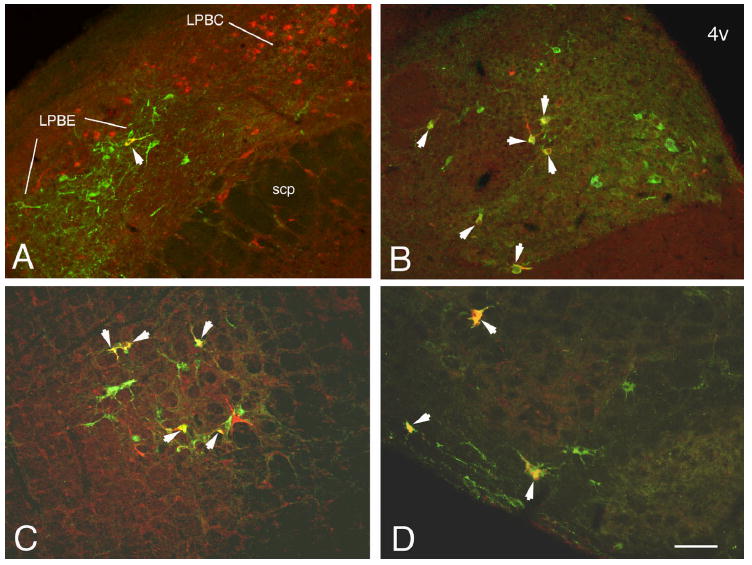
Fluorescent images showing examples of Ctb or PACAP or PACAP and Ctb double-labeled neurons in selected pontine and medullary areas. PACAP neurons show green fluorescence, Ctb labeling shows red, and double-labeled neurons appear yellow/orange (arrows). A: Parabrachial nucleus. B: Nucleus of the solitary tract/dorsal motor nucleus of vagus. C. Ventrolateral medulla. D: Ventral medulla, gigantocellular nucleus. Scale bar = 100 μm.
Within the medulla oblongata, the nucleus of the solitary tract, the DMV, and the VLM contained numerous PACAP/CTb double-labeled neurons. The main localization of retrogradely labeled PACAP neurons in the NTS was in its medial and dorsomedial subnuclei (Figs. 5A, 6B). In the rostral ventrolateral medulla (at –12.0 mm from Bregma), typically at least 50% of detectable PACAP neurons were also CTb positive (Figs. 5C, 6A, Table 1). These double-labeled neurons were localized within the C1 adrenergic cell group area just ventral to the Bötzinger complex and also in the lateral paragigantocellular nucleus (Figs. 5C,D,6A). Double-labeled neurons were observed in the C3 cell group area (Fig. 6A) and more caudally in the C1/A1 cell group and the ventral portion of the gigantocellular nucleus adjacent to the lateral reticular nucleus (Figs. 5D, 6B). By simultaneous triple immunolabeling for PACAP, CTb, and PNMT, several PACAP/CTb neurons in the C1 cell group were also PNMT immunopositive, indicating the likely adrenergic nature of these neurons (Fig. 7). On the other hand, the percentages of retrogradely labeled PACAP-immunopositive neurons showed a decreasing tendency from rostral levels toward the caudal ventrolateral medulla, especially below the transition between C1 and A1 cell groups (Table 1). Likewise, in the NTS and DMV, retrogradely labeled PACAP neurons were more frequently found at the level of the opening of the fourth ventricle at –13.3 mm from Bregma (Fig. 5B) than in more caudal portions of these nuclei (Table 1). Table 1 summarizes relative contributions of sources of PACAP projections to the PVN obtained at distinct rostrocaudal levels of the rat brain from the hypothalamic preoptic area to the caudal brainstem.
Fig. 6.
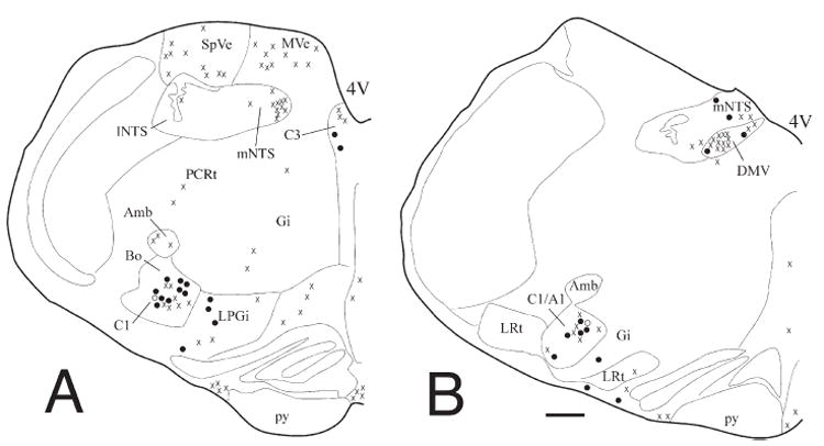
Schematic maps of the distribution of CTb, PACAP, or CTb/PACAP double-labeled neurons in representative levels of the medulla ipsilateral to the injection site. A: Rostral medulla (Bregma –12.0 mm). B: Midcaudal medulla (Bregma –13.3 mm). Symbols: ×, PACAP only; open circles, CTb only; solid circles, PACAP/CTb double-labeled neurons. Each symbol represents a single neuron. Scale bar = 400 μm.
Fig. 7.
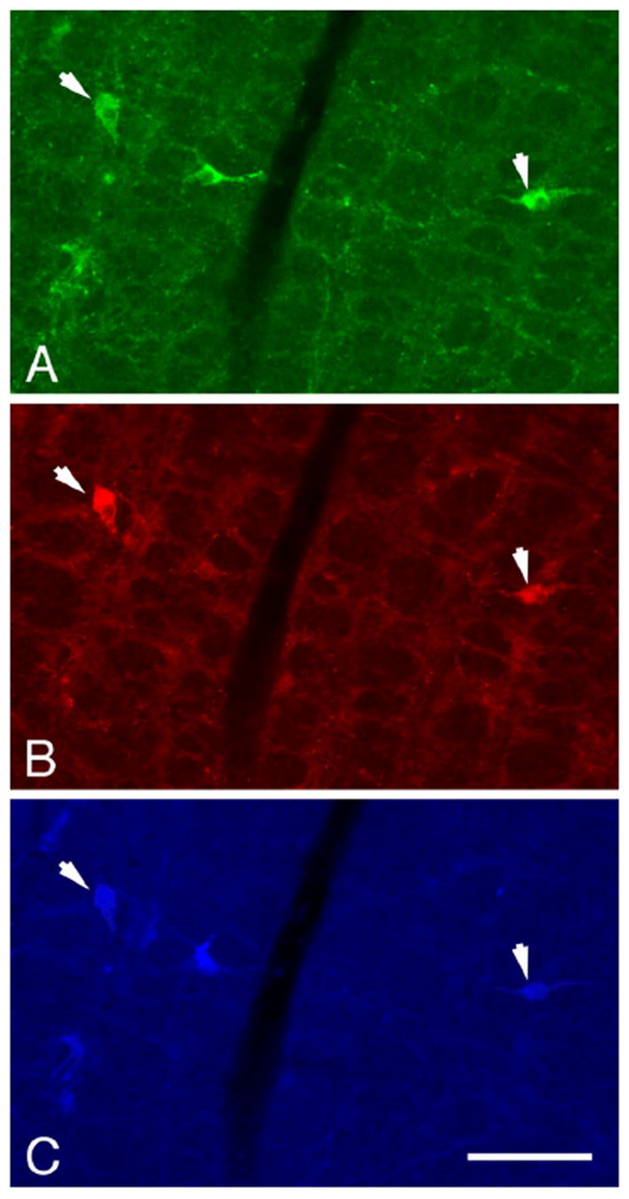
Fluorescent images showing examples of PACAP-, CTb-, and PNMT-labeled neurons in the rostral ventrolateral medulla. A: PACAP. B: CTb. C: PNMT immunolabeling. PACAP neurons show green fluorescence, CTb-labeled neurons show red, and PNMT-labeled neurons are indicated by blue fluorescence. Neurons simultaneously labeled with PACAP, CTb, and PNMT antibodies (triple-labeled neurons) are marked with arrows. Scale bar = 100 μm.
Contralateral contribution
Compared with ipsilateral projections, retrograde labeling was consistently sparser contralateral to the injection site throughout the various PVN-projecting brain areas observed in this study. Furthermore, contralateral CTb labeling was never observed in brain regions that totally lacked ipsilateral contribution. As a result, PACAP/CTb double-labeled neurons on the contralateral side, when present, accounted for only a fraction (approximately 10–20%) of what was found ipsilaterally in the corresponding areas.
DISCUSSION
Intracerebroventricular (icv) or local brain injections of PACAP induce a range of autonomic, behavioral, and neuroendocrine responses. In rodents, icv or intrahypothalamic PACAP injections produce a reduction in food intake (Morley et al., 1992; Chance et al., 1995), a finding recently replicated in the chicken brain, which was inhibited by coadministration of a CRH receptor antagonist, further suggesting the significance of CRH receptors in the anorexic effects of central PACAP administration (Tachibana et al., 2004). PACAP icv also increases plasma vasopressin concentration and mean arterial blood pressure in rats (Murase et al., 1993). Recent functional observations from our laboratory have suggested that PACAP may be an excitatory neuropeptide at the level of the PVN, insofar as intra-PVN microinjection of PACAP increased restraint stress-induced body grooming and reduced locomotor activity in rats (Norrholm et al., 2005) and icv PACAP administration stimulated phosphorylation of the transcription factor CREB in PVN CRH neurons and simultaneously elevated plasma corticosterone levels in rats (Agarwal et al., 2005).
We have proposed that endogenous PACAP nerve fibers innervating various types of PVN neurons (Legradi et al., 1997, 1998) coupled with the presence of specific PACAP receptors in the PVN (Hashimoto et al., 1996) may represent a physiologic background for many of the pharmacologic effects of PACAP administration as summarized above. Because interpretation of functional studies would greatly benefit from the precise understanding of the distribution of PVN-projecting PACAP neurons, the present study utilized intra-PVN injections of the sensitive neuro-anatomical tracer CTb, combined with immunohistochemical identification of retrogradely labeled neurons and PACAP immunoreactivity in the rat hypothalamus and brainstem. In summary, we identified a widespread, multisite origin of PACAP innervation to the PVN from hypothalamic, midbrain, and medullary sources, which suggests the importance of PACAP in homeostatic control mechanisms. These observations provide a strong chemical neuroanatomical foundation for interaction between PACAP and its potential target neurons in the PVN. Targets of PACAPergic innervation thus include neuroendocrine CRH neurons, which control physiologic responses to stressful challenges, but other types of parvo- and magno-cellular neurons, such as those expressing TRH, vasopressin, oxytocin, nitric oxide, glutamate, enkephalin, neuro-tensin, and galanin, should also be considered (Sawchenko et al., 1984; Meister et al., 1990; Hatakeyama et al., 1996; Hrabovszky et al., 2005). In addition, PACAP-containing innervation, originating from the several potential sites identified in the present study, may reach preautonomic projection neurons of the PVN (Swanson and Sawchenko, 1980; Hosoya et al., 1991), which in turn regulate sympathetic and parasympathetic functions by their descending projections (Darlington et al., 1989; Coote et al., 1998).
Specific distribution of PVN-projecting PACAP neurons in the hypothalamus
We found that relatively high numbers of PACAP neurons project to the PVN from several important hypothalamic areas and nuclei. The median and medial preoptic nuclei, medial preoptic area, caudal part of the arcuate nucleus, DMN, VMN, and lateral hypothalamic area contained significant populations of PVN-projecting PACAP neurons. Medium-density projections appeared to originate from the ventrolateral preoptic nucleus, midlevel of DMN, and supramammillary nucleus.
Experimental animal studies have shown that DMN participates in a wide variety of homeostatic, behavioral, and autonomic functions, including feeding and body weight regulation (Bellinger and Mendel, 1978; Bellinger et al., 1979) and cardiovascular control (Bernardis and Bellinger, 1998). It has been shown that DMN neurons mediate stress-induced cardiovascular changes by a balancing mechanism between tonic activity of the inhibitory GABAA and excitatory amino acid receptors (Stotz-Potter et al., 1996). Although it is not known whether PACAP innervation, specifically from the DMN to the PVN, is excitatory or inhibitory in nature, it is conceivable that PACAPergic projections from the DMN modulate functions of PVN neurons. Another hypothalamic nucleus sending major PACAP projections to the PVN is the VMN, which is known to play important roles in feeding, aggression, sexual behavior, and gonadotropin secretion (Matsu-moto and Arai, 1983; Elias et al., 2000), and VMN lesion causes hyperactivity of the hypothalamic-pituitary-adrenal (HPA) axis (Suemaru et al., 1995). The present study also identified PACAP projections from the arcuate nucleus to the PVN whose function is unclear, but it has been suggested that a coordinated and balanced input from arcuate nucleus, DMN, and VMN is required to regulate the activity of the HPA axis (Choi et al., 1996; Bell et al., 2000). It is also possible that PVN-projecting PACAP neurons in the arcuate nucleus, especially in its lateral division, form a subpopulation, if they colocalize with proopiomelanocortin-derived peptides and cocaine- and amphetamine-regulated transcript, which in turn participate in the central regulation of the thyroid and adrenal axes and regulation of food intake (Fekete et al., 2000; Elmquist, 2001; Wittmann et al., 2005).
The lateral hypothalamic area, which is known to send axonal projections to the PVN (Larsen et al., 1994), is thought to be an important regulatory area of feeding, drinking, metabolism, and behavioral activity (Bernardis and Bellinger, 1993). In addition, significant numbers of substance P-containing neurons were identified in the tuberal part of LH, which project to the neuroendocrine subdivisions of the PVN and thus could be involved in the regulation of CRH secretion (Jessop et al., 1992). Similarly, in the present study, we found PVN-projecting PACAP neurons in the rostral tuberal LH, raising the possibility that PACAPergic mechanisms originating from the LH may contribute to the regulation of the neuroendocrine functions of the PVN. The anterior hypothalamic and preoptic areas mediate responses to heat stress (Tanaka et al., 1986) and elicit poststress facilitation of the arterial baroreflex, which may contribute to energy restoration (Nosaka, 1996), whereas the median preoptic nucleus is involved in the central regulation of blood pressure and water and salt homeostasis (Spitznagel et al., 2001). Collectively, the presence of PACAP-containing neurons sending afferents to the PVN from these hypothalamic areas strongly suggests that PACAP neurons may participate in processing of autonomic and neuroendocrine mechanisms at the level of the PVN, presumably acting through magno- and parvocellular vasopressin and parvocellular CRH neurons.
At more caudal, mammillary levels of the hypothalamus, several PACAP neurons contained retrograde labeling from the PVN in the medial part of the supramammillary nucleus (SuMM). The supramammillary nucleus is considered as a bridge between the hippocampus and the amygdala and/or midbrain central gray by creating a route for cognitive–emotional interactions (Pan and Mc-Naughton, 2002). Induction of Fos immunoreactivity was observed in the SuMM in rats after conditioned fear (Beck and Fibiger, 1995) or cold or warm exposure (Kiyohara et al., 1995), as well as after swim stress (Cullinan et al., 1996), and supramammillary PACAP innervation to the PVN may indicate a role of PACAP in mediating emotional and physiological responses. It should also be noted that, under certain circumstances, such as colchicine treatment or endotoxin administration, local parvocellular PVN neurons may synthesize PACAP, and some of them contain CRH immunoreactivity (Hannibal et al., 1995, 1999), so their local axonal arborization in the PVN could be predicted based on previous findings (Liposits et al., 1985). However, current the methodology using retrograde tracer injections within the PVN is unable to resolve neuroanatomical connections between such tightly placed neuronal groups, insofar as both the origin and the target of this type of innervation would be covered by the dense core of the injection site.
Specific distribution of PVN-projecting PACAP neurons in the brainstem
Within the brainstem, PACAP projections to the PVN were found to originate from the PAG, ventrolateral medulla (rostral and midcaudal levels), and medial subnucleus of NTS. The rostral portion of the DMV appeared to send medium-density projections to the PVN. For the midbrain, we identified PACAP projections to the PVN from the ventrolateral PAG, which may be involved in responses to nociception and poststress facilitation of baroreceptor reflexes (Nosaka, 1996) either directly or indirectly, such that a broader role for PACAP in the coordination of defensive behavioral and autonomic output might also be speculated. Activation of the ventrolateral PAG column leads to a hyporeactive pattern of autonomic and behavioral parameters, not unlike manifestations of inescapable stress (Bandler and Shipley, 1994; Depaulis et al., 1994; Keay and Bandler, 2001). Inescapable pain, hemorrhage, and hypotension are paralleled by increases in hypophysiotropic CRH release and/or plasma ACTH and vasopressin levels as well as ventrolateral PAG activation (Plotsky and Vale, 1984; Nakane et al., 1985; Tas-sorelli and Joseph, 1995; Keay and Bandler, 2002). Physiologically identified depressor regions of the caudal ventrolateral PAG were found to project to several subdivisions of the rat PVN (Floyd et al., 1996), but they appear to be significantly more caudal than the PVN-projecting PACAP neurons of our study, suggesting that these PACAP neurons constitute a distinct population of cells. On the other hand, anterograde labeling experiments have shown innervation of the PVN from the rostral ventrolateral PAG (Cameron et al., 1995), an area that shows a closer match with the localization of PVN-projecting PACAP neurons in the present study. Nevertheless, the specific functional characterization of these neurons remains to be established.
Substantial PACAP projections to the PVN were seen originating from the ventrolateral medulla and the NTS/ DMV complex of the dorsal medulla. Earlier chemical neuroanatomical data have shown that the parvocellular PVN receives substantial innervation from medullary C1, C2, and C3 catecholamine cell groups (Cunningham et al., 1990) and sparse innervation from A2 and A6 cell groups (to ventral and periventricular PVN regions; Cunningham and Sawchenko, 1988). Particularly, medial parvocellular CRH neurons receive projections from the C1 neurons of the rostral ventrolateral medulla (Cunningham et al., 1990). Innervation of the magnocellular vasopressinergic PVN comes mainly from the more caudally positioned A1 cell groups of the ventrolateral medulla (Cunningham and Sawchenko, 1988). These regions are known to become activated with a variety of stressors, such as intravenous interleukin-13 (Ericsson et al., 1994), intraperitoneal hypertonic saline (Larsen and Mikkelsen, 1995), and foot shock (Li et al., 1996). Excitatory roles of the ascending catecholaminergic bundle from the medulla to PVN have been implicated in stress-induced increases in plasma ACTH, through activation of parvocellular hypophysiotropic CRH neurons (Weidenfeld and Feldman, 1991; Ericsson et al., 1994). In the present study, we provide further chemical neuroanatomical evidence that a population of the PVN-projecting PACAP neurons in the ventrolateral medulla might also be catecholaminergic, insofar as they colocalized with the adrenergic marker PNMT, confirming and extending our previous observation of PACAP/PNMT-cocontaining innervation of parvocellular PVN neurons (Legradi et al., 1997). Based on our current findings, there may be a relatively higher contribution of the more rostrally placed C1 and the transitional C1/A1 catecholamine cell groups to the PACAP-containing innervation of the PVN, suggesting a more robust innervation to medial parvocellular and preautonomic areas of the PVN compared with the innervation to the magnocellular division.
The ventrolateral medulla has been implicated in regulation of the cardiovascular and respiratory functions (Guyenet et al., 1996). PVN-projecting neurons in the RVLM express Fos immunoreactivity in response to intravenous lypopolysaccharide infusion (Elmquist and Saper, 1996). The same group of neurons is also activated by cardiovascular challenges, including hemorrhage-induced or isovolemic hypotension (Chan and Sawchenko, 1994). In our study, the anatomical localization of the PACAP-containing PVN-projecting neurons in the RVLM suggests that PACAP may participate in cardiovascular regulation through the PVN, perhaps by the activation of vasopressin neurons or through other classes of autonomic-related neurons within the PVN. It is also noteworthy that the non-PVN-projecting population of PACAP neurons in the RVLM may send direct projections to sympathetic preganglionic neurons of the spinal cord, implying a more complex role for PACAP in central autonomic regulation (Legradi et al., 1994b; Dun et al., 1996).
In addition, cold-restraint stress that is associated with gastric lesions has produced an activational pattern in the NTS (Bonaz and Tache, 1994). The presence of PACAP in the PVN-projecting NTS neurons is significant, in that this nucleus receives a variety of cardiovascular, pulmonary, and gastrointestinal afferents and sends information through ascending projections to forebrain target sites such as the PVN, where the signals are further integrated to provide visceromotor and neuroendocrine responses to interoceptive stressors and homeostatic challenges (Buller et al., 2003). Although the specific functional contribution of PVN-projecting PACAP neurons of the medulla oblongata still has to be identified at the cellular level, there is a close distributional match between PACAP/CTb neurons and the localization of stress-activated neurons as mentioned above, suggesting a role for PACAP in autonomic and neuroendocrine manifestations of the response to a number stressors.
Methodological considerations
Immunolabeling for PACAP has been notoriously difficult. Early immunohistochemical studies on the distribution of PACAP were hindered by relatively low sensitivity (Koves et al., 1990,1991; Kivipelto et al., 1992). Fixatives containing acrolein appeared highly beneficial for immunolabeling of various peptides in the brain (King et al., 1983), including PACAP (Legradi et al., 1994a, 1997, 1998). Colchicine pretreatment further enhanced perikaryal visualization of PACAP in a variety of fore-brain and brainstem nuclei (Legradi et al., 1994a, 1997; Hannibal et al., 1995; Hannibal, 2002). In addition to colchicine’s well-known effect of blocking axonal transport, leading to accumulation of peptides in perikarya, high-dose colchicine treatment may induce elevated expression of the PACAP gene in the hypothalamic PVN itself as well as the pontine locus coeruleus and the medullary C1, C2, and C3 adrenergic cell groups (Hannibal et al., 1995; Hannibal, 2002). While keeping these considerations in mind, we would have preferred the combination of high doses of intracerebroventricular colchicine (150–200 μg) followed by acrolein fixation for the current immunohistochemical study, based on the success of earlier studies (Legradi et al., 1994b,1997). However, our pilot experiments had shown that acrolein fixation diminished the detectability of transported CTb, so here we reverted to the use of paraformaldehyde. The masking of epitopes by cross-linking appears to pose a barrier to successful immunodetection of peptides in formaldehyde-fixed tissues (Puchtler and Meloan, 1985). Thus, the inherent low sensitivity of PACAP immunolabeling in paraformaldehyde-fixed tissue was overcome by an antigen-unmasking method: heating the free-floating sections in 10 mM sodium citrate, pH 8.5, at 80°C (Jiao et al., 1999). Heat-induced antigen retrieval provided reliable findings for simultaneous detection of PACAP and CTb immunolabeling, allowing the use of a commercial rabbit anti-PACAP serum for high-sensitivity detection of PACAP immunoreactivity in paraformaldehyde-fixed material. In the present study, 15 minutes of heating time gave optimal antigen retrieval for PACAP without compromising detectability of CTb and minimized heat-induced wrinkling of sections and mechanical damage to tolerable levels. The only exception to this was the subfornical organ, which was distorted by the rigors of processing and whose possible PACAP projections (Piggins et al., 1996; Hannibal, 2002), therefore, will require a separate follow-up study.
Data from the present study were in agreement with the pattern of PACAP immunolabeling observed with the use of a well-characterized monoclonal antibody combined with the biotinylated tyramide amplification system (Hannibal, 2002). Likewise, the high signal-to-noise ratio of the current PACAP immunolabeling product approached the level achieved by either tyramide amplification (Hannibal, 2002) or acrolein fixation (Legradi et al., 1994a, 1997). The possible artifactual effects (e.g., high background) of heat-induced antigen unmasking were successfully avoided by careful monitoring of heating conditions and mandatory use of nonfat dry milk and normal horse serum treatments following section heating as described in Materials and Methods.
Among the brain regions that contained PVN-projecting PACAP neurons, the medullary C1, C2, and C3 cell groups (PNMT-positive, adrenergic neurons) represent a population whose PACAP expression might have been induced to a supraphysiologic level by the high-dose colchicine treatment in the current study. However, the presence of colocalizing PACAP/PNMT-positive nerve terminals at the level of the PVN in close contact with parvocellular TRH (Legradi et al., 1997) or CRH neurons (our unpublished observations), which is evident in low-dose colchicine-injected or untreated material, indicates the existence of axonal projections from putative adrenergic PACAP neurons even under baseline conditions. The finding of PACAP-immunoreactive perikarya in the arcuate nucleus in the present study is consistent with immunohistochemical and in situ hybridization data from two publications (Murase et al., 1995; Piggins et al., 1996), but a more recent study noted the absence of cell body labeling in this nucleus, although one of its published low-magnification photographs appears to show evidence of arcuate nucleus PACAP mRNA expression (Hannibal, 2002). One of the earlier reports also argues that PACAP mRNA in the arcuate nucleus can be evoked by water deprivation (Murase et al., 1995). That PACAP gene expression is induced by colchicine treatment in the arcuate nucleus (either by colchicine’s direct effect or secondarily by the inhibition of water intake) is a possibility, requiring careful further investigation.
Acknowledgments
The skillful technical assistance of Ms. E. Alice Mitchell is greatly appreciated.
Abbreviations
- 3V
third cerebral ventricle
- 4V
fourth cerebral ventricle
- A1
noradrenergic cell group 1
- A2
noradrenergic cell group 2
- A6
noradrenergic cell group 6
- ac
anterior commissure
- AH
anterior hypothalamic area
- Amb
nucleus ambiguous
- aq
cerebral aqueduct
- Arc
arcuate nucleus
- Bo
Bötzinger complex
- C1
catecholaminergic cell group 1
- C2
catecholaminergic cell group 2
- C3
catecholaminergic cell group 3
- CRH
corticotropin-releasing hormone
- CTb
cholera toxin B subunit
- DMN
dorsomedial hypothalamic nucleus
- DMV
dorsal motor nucleus of the vagus
- f
fornix
- Gi
gigantocellular nucleus
- LH
lateral hypothalamic area
- lNTS
nucleus of the solitary tract lateral subnucleus
- LPB
lateral parabrachial nucleus
- LPBC
lateral parabrachial nucleus external subdivision
- LPBE
lateral parabrachial nucleus central subdivision
- LPGi
lateral paragigantocellular nucleus
- LRt
lateral reticular nucleus
- MnPO
median preoptic nucleus
- mNTS
nucleus of the solitary tract medial subnucleus
- MPA
medial preoptic area
- MPBE
lateral parabrachial nucleus medial subdivision
- MPO
medial preoptic nucleus
- MVe
medial vestibular nucleus
- NTS
nucleus of the solitary tract
- och
optic chiasm
- opt
optic tract
- PACAP
pituitary adenylate cyclase-activating polypeptide
- PaDC
paraventricular hypothalamic nucleus, dorsal cap
- PAG
midbrain periaqueductal gray
- PaLM
paraventricular hypothalamic nucleus, lateral magnocellular
- PaMP
paraventricular hypothalamic nucleus, medial parvocellular
- PBN
parabrachial nucleus
- PCRt
parvocellular reticular nucleus
- PeF
perifornical nucleus
- PeH
perifornical hypothalamus
- PH
posterior hypothalamus
- PS
parastrial nucleus
- PVG
periventricular gray
- PVN
paraventricular hypothalamic nucleus
- py
pyramidal tract
- RCh
retrochiasmatic nucleus
- RVLM
rostral ventrolateral medulla
- Sch
suprachiasmatic nucleus
- Scp
superior cerebellar peduncle
- Shy
septohypothalamic nucleus
- SpVe
spinal vestibular nucleus
- STMAL
bed nucleus of the stria terminalis, lateral division, anterolateral part
- STMAM
bed nucleus of the stria terminalis, medial division, anteromedial part
- SuMM
supramammillary nucleus medial subnucleus
- VLM
ventrolateral medulla
- VMN
ventromedial hypothalamic nucleus
- VMPO
ventromedial preoptic nucleus
Footnotes
Grant sponsor: National Institutes of Health; Grant number: R01 MH63320.
LITERATURE CITED
- Agarwal A, Halvorson LM, Legradi G. Pituitary adenylate cyclase-activating polypeptide (PACAP) mimics neuroendocrine and behavioral manifestations of stress: evidence for PKA-mediated expression of the corticotropin-releasing hormone (CRH) gene. Brain Res Mol Brain Res. 2005;138:45–57. doi: 10.1016/j.molbrainres.2005.03.016. [DOI] [PMC free article] [PubMed] [Google Scholar]
- Arimura A. Perspectives on pituitary adenylate cyclase activating polypeptide (PACAP) in the neuroendocrine, endocrine, and nervous systems. Jpn J Physiol. 1998;48:301–331. doi: 10.2170/jjphysiol.48.301. [DOI] [PubMed] [Google Scholar]
- Arimura A, Somogyvari-Vigh A, Miyata A, Mizuno K, Coy DH, Kitada C. Tissue distribution of PACAP as determined by RIA: highly abundant in the rat brain and testes. Endocrinology. 1991;129:2787–2789. doi: 10.1210/endo-129-5-2787. [DOI] [PubMed] [Google Scholar]
- Bandler R, Shipley MT. Columnar organization in the midbrain periaqueductal gray: modules for emotional expression? Trends Neurosci. 1994;17:379–389. doi: 10.1016/0166-2236(94)90047-7. [DOI] [PubMed] [Google Scholar]
- Beck CH, Fibiger HC. Conditioned fear-induced changes in behavior and in the expression of the immediate early gene c-fos: with and without diazepam pretreatment. J Neurosci. 1995;15:709–720. doi: 10.1523/JNEUROSCI.15-01-00709.1995. [DOI] [PMC free article] [PubMed] [Google Scholar]
- Bell ME, Bhatnagar S, Akana SF, Choi S, Dallman MF. Disruption of arcuate/paraventricular nucleus connections changes body energy balance and response to acute stress. J Neurosci. 2000;20:6707–6713. doi: 10.1523/JNEUROSCI.20-17-06707.2000. [DOI] [PMC free article] [PubMed] [Google Scholar]
- Bellinger LL, Mendel VE. Hypophysectomy alters the diurnal food intake patterns in rats. Proc Soc Exp Biol Med. 1978;159:80–83. doi: 10.3181/00379727-159-40288. [DOI] [PubMed] [Google Scholar]
- Bellinger LL, Bernardis LL, Brooks S. The effect of dorsomedial hypothalamic nuclei lesions on body weight regulation. Neuroscience. 1979;4:659–665. doi: 10.1016/0306-4522(79)90142-8. [DOI] [PubMed] [Google Scholar]
- Bernardis LL, Bellinger LL. The lateral hypothalamic area revisited: neuroanatomy, body weight regulation, neuroendocrinology and metabolism. Neurosci Biobehav Rev. 1993;17:141–193. doi: 10.1016/s0149-7634(05)80149-6. [DOI] [PubMed] [Google Scholar]
- Bernardis LL, Bellinger LL. The dorsomedial hypothalamic nucleus revisited: 1998 update. Proc Soc Exp Biol Med. 1998;218:284–306. doi: 10.3181/00379727-218-44296. [DOI] [PubMed] [Google Scholar]
- Bonaz B, Tache Y. Induction of Fos immunoreactivity in the rat brain after cold-restraint induced gastric lesions and fecal excretion. Brain Res. 1994;652:56–64. doi: 10.1016/0006-8993(94)90316-6. [DOI] [PubMed] [Google Scholar]
- Buller KM, Dayas CV, Day TA. Descending pathways from the paraventricular nucleus contribute to the recruitment of brainstem nuclei following a systemic immune challenge. Neuroscience. 2003;118:189–203. doi: 10.1016/s0306-4522(02)00808-4. [DOI] [PubMed] [Google Scholar]
- Cameron AA, Khan IA, Westlund KN, Cliffer KD, Willis WD. The efferent projections of the periaqueductal gray in the rat: a Phaseolus vulgaris-leucoagglutinin study. I. Ascending projections. J Comp Neurol. 1995;351:568–584. doi: 10.1002/cne.903510407. [DOI] [PubMed] [Google Scholar]
- Chan RK, Sawchenko PE. Spatially and temporally differentiated patterns of c-fos expression in brainstem catecholaminergic cell groups induced by cardiovascular challenges in the rat. J Comp Neurol. 1994;348:433–460. doi: 10.1002/cne.903480309. [DOI] [PubMed] [Google Scholar]
- Chance WT, Thompson H, Thomas I, Fischer JE. Anorectic and neurochemical effects of pituitary adenylate cyclase activating polypeptide in rats. Peptides. 1995;16:1511–1516. doi: 10.1016/0196-9781(95)02048-9. [DOI] [PubMed] [Google Scholar]
- Choi S, Horsley C, Aguila S, Dallman MF. The hypothalamic ventromedial nuclei couple activity in the hypothalamo-pituitary-adrenal axis to the morning fed or fasted state. J Neurosci. 1996;16:8170–8180. doi: 10.1523/JNEUROSCI.16-24-08170.1996. [DOI] [PMC free article] [PubMed] [Google Scholar]
- Coote JH, Yang Z, Pyner S, Deering J. Control of sympathetic outflows by the hypothalamic paraventricular nucleus. Clin Exp Pharmacol Physiol. 1998;25:461–463. doi: 10.1111/j.1440-1681.1998.tb02235.x. [DOI] [PubMed] [Google Scholar]
- Cullinan WE, Helmreich DL, Watson SJ. Fos expression in forebrain afferents to the hypothalamic paraventricular nucleus following swim stress. J Comp Neurol. 1996;368:88–99. doi: 10.1002/(SICI)1096-9861(19960422)368:1<88::AID-CNE6>3.0.CO;2-G. [DOI] [PubMed] [Google Scholar]
- Cunningham ET, Jr, Sawchenko PE. Anatomical specificity of nor-adrenergic inputs to the paraventricular and supraoptic nuclei of the rat hypothalamus. J Comp Neurol. 1988;274:60–76. doi: 10.1002/cne.902740107. [DOI] [PubMed] [Google Scholar]
- Cunningham ET, Jr, Bohn MC, Sawchenko PE. Organization of adrenergic inputs to the paraventricular and supraoptic nuclei of the hypothalamus in the rat. J Comp Neurol. 1990;292:651– 667. doi: 10.1002/cne.902920413. [DOI] [PubMed] [Google Scholar]
- Darlington DN, Miyamoto M, Keil LC, Dallman MF. Paraventricular stimulation with glutamate elicits bradycardia and pituitary responses. Am J Physiol. 1989;256:R112–R119. doi: 10.1152/ajpregu.1989.256.1.R112. [DOI] [PubMed] [Google Scholar]
- Depaulis A, Keay KA, Bandler R. Quiescence and hyporeactivity evoked by activation of cell bodies in the ventrolateral midbrain peri-aqueductal gray of the rat. Exp Brain Res. 1994;99:75–83. doi: 10.1007/BF00241413. [DOI] [PubMed] [Google Scholar]
- Dun NJ, Miyazaki T, Tang H, Dun EC. Pituitary adenylate cyclase activating polypeptide immunoreactivity in the rat spinal cord and medulla: implication of sensory and autonomic functions. Neuroscience. 1996;73:677–686. doi: 10.1016/0306-4522(96)00057-7. [DOI] [PubMed] [Google Scholar]
- Elias CF, Kelly JF, Lee CE, Ahima RS, Drucker DJ, Saper CB, Elmquist JK. Chemical characterization of leptin-activated neurons in the rat brain. J Comp Neurol. 2000;423:261–281. [PubMed] [Google Scholar]
- Elmquist JK. Hypothalamic pathways underlying the endocrine, autonomic, and behavioral effects of leptin. Int J Obes Relat Metab Disord. 2001;25(Suppl 5):S78–S82. doi: 10.1038/sj.ijo.0801918. [DOI] [PubMed] [Google Scholar]
- Elmquist JK, Saper CB. Activation of neurons projecting to the paraventricular hypothalamic nucleus by intravenous lipopolysaccharide. J Comp Neurol. 1996;374:315–331. doi: 10.1002/(SICI)1096-9861(19961021)374:3<315::AID-CNE1>3.0.CO;2-4. [DOI] [PubMed] [Google Scholar]
- Ericsson A, Kovacs KJ, Sawchenko PE. A functional anatomical analysis of central pathways subserving the effects of interleukin-1 on stress-related neuroendocrine neurons. J Neurosci. 1994;14:897–913. doi: 10.1523/JNEUROSCI.14-02-00897.1994. [DOI] [PMC free article] [PubMed] [Google Scholar]
- Fekete C, Legradi G, Mihaly E, Huang QH, Tatro JB, Rand WM, Emerson CH, Lechan RM. Alpha-melanocyte-stimulating hormone is contained in nerve terminals innervating thyrotropin-releasing hormone-synthesizing neurons in the hypothalamic paraventricular nucleus and prevents fasting-induced suppression of prothyrotropin-releasing hormone gene expression. J Neurosci. 2000;20:1550–1558. doi: 10.1523/JNEUROSCI.20-04-01550.2000. [DOI] [PMC free article] [PubMed] [Google Scholar]
- Floyd NS, Keay KA, Arias CM, Sawchenko PE, Bandler R. Projections from the ventrolateral periaqueductal gray to endocrine regulatory subdivisions of the paraventricular nucleus of the hypothalamus in the rat. Neurosci Lett. 1996;220:105–108. doi: 10.1016/s0304-3940(96)13240-7. [DOI] [PubMed] [Google Scholar]
- Guyenet PG, Koshiya N, Huangfu D, Baraban SC, Stornetta RL, Li YW. Role of medulla oblongata in generation of sympathetic and vagal outflows. Prog Brain Res. 1996;107:127–144. doi: 10.1016/s0079-6123(08)61862-2. [DOI] [PubMed] [Google Scholar]
- Hannibal J. Pituitary adenylate cyclase-activating peptide in the rat central nervous system: an immunohistochemical and in situ hybridization study. J Comp Neurol. 2002;453:389–417. doi: 10.1002/cne.10418. [DOI] [PubMed] [Google Scholar]
- Hannibal J, Mikkelsen JD, Fahrenkrug J, Larsen PJ. Pituitary adenylate cyclase-activating peptide gene expression in corticotropin-releasing factor-containing parvicellular neurons of the rat hypothalamic paraventricular nucleus is induced by colchicine, but not by adrenalectomy, acute osmotic, ether, or restraint stress. Endocrinology. 1995;136:4116–4124. doi: 10.1210/endo.136.9.7649120. [DOI] [PubMed] [Google Scholar]
- Hannibal J, Jessop DS, Fahrenkrug J, Harbuz MS, Larsen PJ. PACAP gene expression in neurons of the rat hypothalamo-pituitary-adrenocortical axis is induced by endotoxin and interleukin-1beta. Neuroendocrinology. 1999;70:73–82. doi: 10.1159/000054461. [DOI] [PubMed] [Google Scholar]
- Hashimoto H, Nogi H, Mori K, Ohishi H, Shigemoto R, Yamamoto K, Matsuda T, Mizuno N, Nagata S, Baba A. Distribution of the mRNA for a pituitary adenylate cyclase-activating polypeptide receptor in the rat brain: an in situ hybridization study. J Comp Neurol. 1996;371:567–577. doi: 10.1002/(SICI)1096-9861(19960805)371:4<567::AID-CNE6>3.0.CO;2-2. [DOI] [PubMed] [Google Scholar]
- Hatakeyama S, Kawai Y, Ueyama T, Senba E. Nitric oxide synthase-containing magnocellular neurons of the rat hypothalamus synthesize oxytocin and vasopressin and express Fos following stress stimuli. J Chem Neuroanat. 1996;11:243–256. doi: 10.1016/s0891-0618(96)00166-4. [DOI] [PubMed] [Google Scholar]
- Herman JP, Cullinan WE, Ziegler DR, Tasker JG. Role of the paraventricular nucleus microenvironment in stress integration. Eur J Neurosci. 2002;16:381–385. doi: 10.1046/j.1460-9568.2002.02133.x. [DOI] [PubMed] [Google Scholar]
- Hosoya Y, Sugiura Y, Okado N, Loewy AD, Kohno K. Descending input from the hypothalamic paraventricular nucleus to sympathetic preganglionic neurons in the rat. Exp Brain Res. 1991;85:10–20. doi: 10.1007/BF00229982. [DOI] [PubMed] [Google Scholar]
- Hrabovszky E, Wittmann G, Turi GF, Liposits Z, Fekete C. Hypophysiotropic thyrotropin-releasing hormone and corticotropin-releasing hormone neurons of the rat contain vesicular glutamate transporter-2. Endocrinology. 2005;146:341–347. doi: 10.1210/en.2004-0856. [DOI] [PubMed] [Google Scholar]
- Jessop DS, Chowdrey HS, Larsen PJ, Lightman SL. Substance P: multifunctional peptide in the hypothalamo-pituitary system? J Endocrinol. 1992;132:331–337. doi: 10.1677/joe.0.1320331. [DOI] [PubMed] [Google Scholar]
- Jiao Y, Sun Z, Lee T, Fusco FR, Kimble TD, Meade CA, Cuthbertson S, Reiner A. A simple and sensitive antigen retrieval method for free-floating and slide-mounted tissue sections. J Neurosci Methods. 1999;93:149–162. doi: 10.1016/s0165-0270(99)00142-9. [DOI] [PubMed] [Google Scholar]
- Keay KA, Bandler R. Parallel circuits mediating distinct emotional coping reactions to different types of stress. Neurosci Biobehav Rev. 2001;25:669–678. doi: 10.1016/s0149-7634(01)00049-5. [DOI] [PubMed] [Google Scholar]
- Keay KA, Bandler R. Distinct central representations of inescapable and escapable pain: observations and speculation. Exp Physiol. 2002;87:275–279. doi: 10.1113/eph8702355. [DOI] [PubMed] [Google Scholar]
- King JC, Lechan RM, Kugel G, Anthony EL. Acrolein: a fixative for immunocytochemical localization of peptides in the central nervous system. J Histochem Cytochem. 1983;31:62– 68. doi: 10.1177/31.1.6187805. [DOI] [PubMed] [Google Scholar]
- Kivipelto L, Absood A, Arimura A, Sundler F, Hakanson R, Panula P. The distribution of pituitary adenylate cyclase-activating polypeptide-like immunoreactivity is distinct from helodermin- and helospectin-like immunoreactivities in the rat brain. J Chem Neuroanat. 1992;5:85–94. doi: 10.1016/0891-0618(92)90036-p. [DOI] [PubMed] [Google Scholar]
- Kiyohara T, Miyata S, Nakamura T, Shido O, Nakashima T, Shibata M. Differences in Fos expression in the rat brains between cold and warm ambient exposures. Brain Res Bull. 1995;38:193–201. doi: 10.1016/0361-9230(95)00093-t. [DOI] [PubMed] [Google Scholar]
- Koves K, Arimura A, Somogyvari-Vigh A, Vigh S, Miller J. Immunohistochemical demonstration of a novel hypothalamic peptide, pituitary adenylate cyclase-activating polypeptide, in the ovine hypothalamus. Endocrinology. 1990;127:264–271. doi: 10.1210/endo-127-1-264. [DOI] [PubMed] [Google Scholar]
- Koves K, Arimura A, Gorcs TG, Somogyvari-Vigh A. Comparative distribution of immunoreactive pituitary adenylate cyclase activating polypeptide and vasoactive intestinal polypeptide in rat forebrain. Neuroendocrinology. 1991;54:159–169. doi: 10.1159/000125864. [DOI] [PubMed] [Google Scholar]
- Larsen PJ, Mikkelsen JD. Functional identification of central afferent projections conveying information of acute “stress” to the hypothalamic paraventricular nucleus. J Neurosci. 1995;15:2609–2627. doi: 10.1523/JNEUROSCI.15-04-02609.1995. [DOI] [PMC free article] [PubMed] [Google Scholar]
- Larsen PJ, Hay-Schmidt A, Mikkelsen JD. Efferent connections from the lateral hypothalamic region and the lateral preoptic area to the hypothalamic paraventricular nucleus of the rat. J Comp Neurol. 1994;342:299–319. doi: 10.1002/cne.903420211. [DOI] [PubMed] [Google Scholar]
- Legradi G, Shioda S, Arimura A. Pituitary adenylate cyclase-activating polypeptide-like immunoreactivity in autonomic regulatory areas of the rat medulla oblongata. Neurosci Lett. 1994a;176:193–196. doi: 10.1016/0304-3940(94)90080-9. [DOI] [PubMed] [Google Scholar]
- Legradi G, Shioda S, Takaki A, Arimura A. Immunohistochemical mapping of pituitary adenylate cyclase-activating polypeptide PACAP in the rat brain. Soc Neurosci Abstr. 1994b;20:516. [Google Scholar]
- Legradi G, Hannibal J, Lechan RM. Association between pituitary adenylate cyclase-activating polypeptide and thyrotropin-releasing hormone in the rat hypothalamus. J Chem Neuroanat. 1997;13:265–279. doi: 10.1016/s0891-0618(97)10002-3. [DOI] [PubMed] [Google Scholar]
- Legradi G, Hannibal J, Lechan RM. Pituitary adenylate cyclase-activating polypeptide-nerve terminals densely innervate corticotropin-releasing hormone-neurons in the hypothalamic paraventricular nucleus of the rat. Neurosci Lett. 1998;246:145–148. doi: 10.1016/s0304-3940(98)00255-9. [DOI] [PubMed] [Google Scholar]
- Li HY, Ericsson A, Sawchenko PE. Distinct mechanisms underlie activation of hypothalamic neurosecretory neurons and their medullary catecholaminergic afferents in categorically different stress paradigms. Proc Natl Acad Sci U S A. 1996;93:2359–2364. doi: 10.1073/pnas.93.6.2359. [DOI] [PMC free article] [PubMed] [Google Scholar]
- Liposits Z, Paull WK, Setalo G, Vigh S. Evidence for local cortico-tropin releasing factor (CRF)-immunoreactive neuronal circuits in the paraventricular nucleus of the rat hypothalamus. An electron microscopic immunohistochemical analysis. Histochemistry. 1985;83:5–16. doi: 10.1007/BF00495294. [DOI] [PubMed] [Google Scholar]
- Masuo Y, Suzuki N, Matsumoto H, Tokito F, Matsumoto Y, Tsuda M, Fujino M. Regional distribution of pituitary adenylate cyclase activating polypeptide (PACAP) in the rat central nervous system as determined by sandwich-enzyme immunoassay. Brain Res. 1993;602:57– 63. doi: 10.1016/0006-8993(93)90241-e. [DOI] [PubMed] [Google Scholar]
- Matsumoto A, Arai Y. Sex difference in volume of the ventromedial nucleus of the hypothalamus in the rat. Endocrinol Jpn. 1983;30:277–280. doi: 10.1507/endocrj1954.30.277. [DOI] [PubMed] [Google Scholar]
- McKellar S, Loewy AD. Organization of some brain stem afferents to the paraventricular nucleus of the hypothalamus in the rat. Brain Res. 1981;217:351–357. doi: 10.1016/0006-8993(81)90010-x. [DOI] [PubMed] [Google Scholar]
- Meister B, Villar MJ, Ceccatelli S, Hokfelt T. Localization of chemical messengers in magnocellular neurons of the hypothalamic supraoptic and paraventricular nuclei: an immunohistochemical study using experimental manipulations. Neuroscience. 1990;37:603– 633. doi: 10.1016/0306-4522(90)90094-k. [DOI] [PubMed] [Google Scholar]
- Mihaly E, Legradi G, Fekete C, Lechan RM. Efferent projections of ProTRH neurons in the ventrolateral periaqueductal gray. Brain Res. 2001;919:185–197. doi: 10.1016/s0006-8993(01)02962-6. [DOI] [PubMed] [Google Scholar]
- Miyata A, Arimura A, Dahl RR, Minamino N, Uehara A, Jiang L, Culler MD, Coy DH. Isolation of a novel 38 residue-hypothalamic polypeptide which stimulates adenylate cyclase in pituitary cells. Biochem Biophys Res Commun. 1989;164:567–574. doi: 10.1016/0006-291x(89)91757-9. [DOI] [PubMed] [Google Scholar]
- Moga MM, Saper CB. Neuropeptide-immunoreactive neurons projecting to the paraventricular hypothalamic nucleus in the rat. J Comp Neurol. 1994;346:137–150. doi: 10.1002/cne.903460110. [DOI] [PubMed] [Google Scholar]
- Morley JE, Horowitz M, Morley PM, Flood JF. Pituitary adenylate cyclase activating polypeptide (PACAP) reduces food intake in mice. Peptides. 1992;13:1133–1135. doi: 10.1016/0196-9781(92)90019-y. [DOI] [PubMed] [Google Scholar]
- Murase T, Kondo K, Otake K, Oiso Y. Pituitary adenylate cyclase-activating polypeptide stimulates arginine vasopressin release in conscious rats. Neuroendocrinology. 1993;57:1092–1096. doi: 10.1159/000126475. [DOI] [PubMed] [Google Scholar]
- Murase T, Kondo K, Arima H, Iwasaki Y, Ito M, Miura Y, Oiso Y. The expression of pituitary adenylate cyclase-activating polypeptide (PACAP) mRNA in rat brain: possible role of endogenous PACAP in vasopressin release. Neurosci Lett. 1995;185:103–106. doi: 10.1016/0304-3940(94)11235-b. [DOI] [PubMed] [Google Scholar]
- Nakane T, Audhya T, Kanie N, Hollander CS. Evidence for a role of endogenous corticotropin-releasing factor in cold, ether, immobilization, and traumatic stress. Proc Natl Acad Sci U S A. 1985;82:1247–1251. doi: 10.1073/pnas.82.4.1247. [DOI] [PMC free article] [PubMed] [Google Scholar]
- Norrholm SD, Das M, Legradi G. Behavioral effects of local micro-infusion of pituitary adenylate cyclase activating polypeptide (PACAP) into the paraventricular nucleus of the hypothalamus (PVN) Regul Pept. 2005;128:33–41. doi: 10.1016/j.regpep.2004.12.023. [DOI] [PMC free article] [PubMed] [Google Scholar]
- Nosaka S. Modifications of arterial baroreflexes: obligatory roles in cardiovascular regulation in stress and poststress recovery. Jpn J Physiol. 1996;46:271–288. doi: 10.2170/jjphysiol.46.271. [DOI] [PubMed] [Google Scholar]
- Pan WX, McNaughton N. The role of the medial supramammillary nucleus in the control of hippocampal theta activity and behaviour in rats. Eur J Neurosci. 2002;16:1797–1809. doi: 10.1046/j.1460-9568.2002.02267.x. [DOI] [PubMed] [Google Scholar]
- Paxinos G, Watson C. The rat brain in stereotaxic coordinates. Amsterdam: Elsevier/Academic Press; 2005. [Google Scholar]
- Piggins HD, Stamp JA, Burns J, Rusak B, Semba K. Distribution of pituitary adenylate cyclase activating polypeptide (PACAP) immuno-reactivity in the hypothalamus and extended amygdala of the rat. J Comp Neurol. 1996;376:278–294. doi: 10.1002/(SICI)1096-9861(19961209)376:2<278::AID-CNE9>3.0.CO;2-0. [DOI] [PubMed] [Google Scholar]
- Plotsky PM, Vale W. Hemorrhage-induced secretion of corticotropin-releasing factor-like immunoreactivity into the rat hypophysial portal circulation and its inhibition by glucocorticoids. Endocrinology. 1984;114:164–169. doi: 10.1210/endo-114-1-164. [DOI] [PubMed] [Google Scholar]
- Puchtler H, Meloan SN. On the chemistry of formaldehyde fixation and its effects on immunohistochemical reactions. Histochemistry. 1985;82:201–204. doi: 10.1007/BF00501395. [DOI] [PubMed] [Google Scholar]
- Sawchenko PE, Swanson LW, Vale WW. Corticotropin-releasing factor: co-expression within distinct subsets of oxytocin-, vasopressin-, and neurotensin-immunoreactive neurons in the hypothalamus of the male rat. J Neurosci. 1984;4:1118–1129. doi: 10.1523/JNEUROSCI.04-04-01118.1984. [DOI] [PMC free article] [PubMed] [Google Scholar]
- Sawchenko PE, Brown ER, Chan RK, Ericsson A, Li HY, Roland BL, Kovacs KJ. The paraventricular nucleus of the hypothalamus and the functional neuroanatomy of visceromotor responses to stress. Prog Brain Res. 1996;107:201–222. doi: 10.1016/s0079-6123(08)61866-x. [DOI] [PubMed] [Google Scholar]
- Shioda S, Yada T, Nakajo S, Nakaya K, Nakai Y, Arimura A. Pituitary adenylate cyclase-activating polypeptide (PACAP): a novel regulator of vasopressin-containing neurons. Brain Res. 1997;765:81–90. doi: 10.1016/s0006-8993(97)00512-x. [DOI] [PubMed] [Google Scholar]
- Spitznagel H, Baulmann J, Blume A, Unger T, Culman J. C-FOS expression in the rat brain in response to substance P and neurokinin B. Brain Res. 2001;916:11–21. doi: 10.1016/s0006-8993(01)02858-x. [DOI] [PubMed] [Google Scholar]
- Stotz-Potter EH, Morin SM, DiMicco JA. Effect of microinjection of muscimol into the dorsomedial or paraventricular hypothalamic nucleus on air stress-induced neuroendocrine and cardiovascular changes in rats. Brain Res. 1996;742:219–224. doi: 10.1016/s0006-8993(96)01011-6. [DOI] [PubMed] [Google Scholar]
- Suemaru S, Darlington DN, Akana SF, Cascio CS, Dallman MF. Ventromedial hypothalamic lesions inhibit corticosteroid feedback regulation of basal ACTH during the trough of the circadian rhythm. Neuroendocrinology. 1995;61:453–463. doi: 10.1159/000126868. [DOI] [PubMed] [Google Scholar]
- Swanson LW. Brain maps: structure of the rat brain. Amsterdam: Elsevier; 1998. [Google Scholar]
- Swanson LW, Sawchenko PE. Paraventricular nucleus: a site for the integration of neuroendocrine and autonomic mechanisms. Neuroendocrinology. 1980;31:410–417. doi: 10.1159/000123111. [DOI] [PubMed] [Google Scholar]
- Tachibana T, Saito ES, Takahashi H, Saito S, Tomonaga S, Boswell T, Furuse M. Anorexigenic effects of pituitary adenylate cyclase-activating polypeptide and vasoactive intestinal peptide in the chick brain are mediated by corticotrophin-releasing factor. Regul Pept. 2004;120:99–105. doi: 10.1016/j.regpep.2004.02.016. [DOI] [PubMed] [Google Scholar]
- Tanaka H, Kanosue K, Nakayama T, Shen Z. Grooming, body extension, and vasomotor responses induced by hypothalamic warming at different ambient temperatures in rats. Physiol Behav. 1986;38:145–151. doi: 10.1016/0031-9384(86)90145-9. [DOI] [PubMed] [Google Scholar]
- Tassorelli C, Joseph SA. Systemic nitroglycerin induces Fos immunoreactivity in brainstem and forebrain structures of the rat. Brain Res. 1995;682:167–181. doi: 10.1016/0006-8993(95)00348-t. [DOI] [PubMed] [Google Scholar]
- Weidenfeld J, Feldman S. Effect of hypothalamic norepinephrine depletion on median eminence CRF-41 content and serum ACTH in control and adrenalectomized rats. Brain Res. 1991;542:201–204. doi: 10.1016/0006-8993(91)91567-k. [DOI] [PubMed] [Google Scholar]
- Wittmann G, Liposits Z, Lechan RM, Fekete C. Origin of cocaine- and amphetamine-regulated transcript-containing axons innervating hypophysiotropic corticotropin-releasing hormone-synthesizing neurons in the rat. Endocrinology. 2005;146:2985–2991. doi: 10.1210/en.2005-0178. [DOI] [PubMed] [Google Scholar]