


Figure 3.
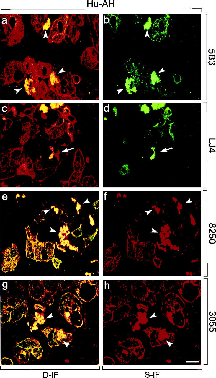
Distribution of phosphorylated CK8 or CK18, and total CK8/18 in human alcoholic hepatitis (Hu-AH). Immunofluorescence double-labeling was performed on frozen tissue sections using antibodies to phospho-CK8 (5B3, LJ4; green in a–d), phospho-CK18 (8250, 3055; red in e–h), and to total CK8/18 (CK antibodies; red in a, c; green in e, g). Double- (D-IF) and corresponding single- (S-IF) label confocal micrographs are shown. 5B3, 8250, 3055: Note the shift of phospho-CK8/18 staining in AH from the cell periphery to the whole cytoplasm with enhanced labeling of the cytokeratin filament network and of MBs. Examples of MBs are indicated by arrowheads. LJ4: In AH, immunoreactivity with cytokeratin filaments and with a subset of MBs is found. Arrows in c and d indicate nonreactive MBs. Bar = 20 μm.