


Figure 4. Predicted mechanisms of action of synthetic riboswitches.
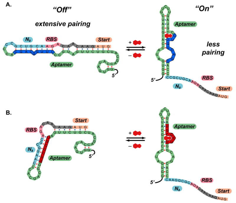
A.) Predicted mechanism of action of clone 8.1. In the absence of theophylline (left), the 5′-region can adopt a highly folded structure that extensively pairs the region that includes ribosome binding site to part of the aptamer sequence (shown in dark blue). In the presence of theophylline (right), the secondary structure shifts, such that the ribosome binding site is exposed. The secondary structure shown for the theophylline aptamer predicted by mFold and is identical to the structure determined by NMR. B.) Predicted mechanism of action of clone 8.2. In this case, the region containing the ribosome binding site pairs to a different region of the aptamer in the “off” state (shown in red). This demonstrates that several motifs may function as synthetic riboswitches.