


Figure 5.
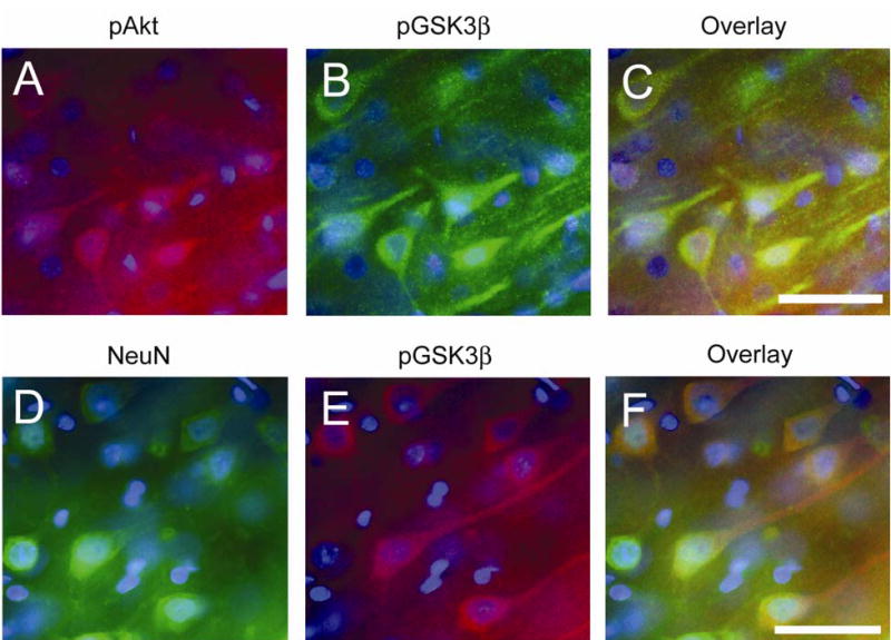
Double immunofluorescent staining of phosphorylation of Akt (pAkt) (red) and of GSK3β (pGSK3β) (green), and NeuN (green) and pGSK3β (red) in the cerebral cortex 6 h after SAH. Nuclei were counterstained with DAPI (blue). pAkt-positive cells (A) and pGSK3β-positive cells. (B) were observed in the cerebral cortex. (C) An overlapped image shows that pAkt-positive cells colocalized with pGSK3β-positive cells. (D) NeuN-positive cells reveal neurons in the cerebral cortex. (E) pGSK3β immunohistochemistry shows pGSK3β-positive cells in the same view. (F) An overlapped image shows that pGSK3β-positive cells colocalized with neurons. Scale bars represent 50 μm.