


Figure 2.
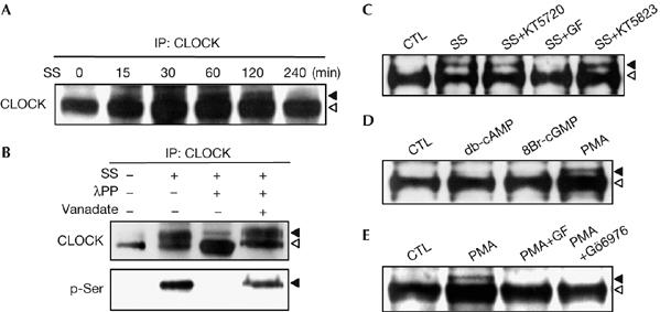
Protein kinase C-dependent phosphorylation of CLOCK. (A) Time-course changes of the electrophoretic mobility of CLOCK. CLOCK immunoprecipitates (IP) were prepared at the indicated time points after serum shock (SS), separated by long SDS–PAGE on 6% gels and visualized by immunoblotting with anti-CLOCK. Arrowheads indicate the slow-migrating band (black) and the fast-migrating band (white). (B) CLOCK immunoprecipitates from the cells before and after serum shock (30 min) were incubated with λ protein phosphatase (λPP) in the absence or presence of vanadate (Na3VO4) and were assessed by immunoblotting with the indicated antibodies. (C) The effect of various kinase inhibitors on CLOCK phosphorylation induced by serum shock. Cells were treated with 0.5 μM KT5720, 5 μM GF109203X (GF) or 1 μM KT5823 1 h before serum shock. Arrowheads indicate the phosphorylated form (black) and the naked form (white). (D) The effect of diverse kinase activators on phosphorylation of CLOCK. Cells were treated with 1 mM db-cAMP, 1 mM 8Br-cGMP or 1 μM PMA for 30 min and whole-cell lysates were subjected to immunoblotting with anti-CLOCK. (E) Characterization of PMA-induced CLOCK phosphorylation. Cells were treated with 5 μM GF109203X (GF) or 5 μM Gö6976 1 h before incubation with PMA for 30 min and analysed as described in (D). CTL, control; PMA, phorbol myristic acetate; SDS–PAGE, sodium dodecyl sulphate–polyacrylamide gel electrophoresis.