


Figure 9.
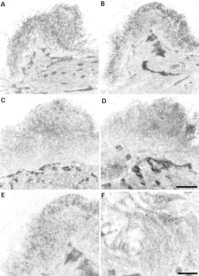
Immunogold stainings for Aβ40 and Aβ42. A and B: Serial sections showing a vascular amyloid deposit staining for Aβ40 (A) and Aβ42 (B). C and D: Amyloid focus staining for Aβ42 (D), but not for Aβ40 (C). The existence of foci that contain Aβ42, but not Aβ40 suggests that Aβ42 is the initially deposited form. A and C and B and D are from the same ultrathin section. E: Higher magnification of B. The amyloid deposit consists of randomly oriented slightly curved fibers with a thickness of 8 to 10 nm. F: In comparison, amyloid fibers in plaques are straight and form rigid bundles (immunogold for Aβ42). Scale bars, 1 μm (A–D) and 0.5 μm (E and F).