


Figure 10.
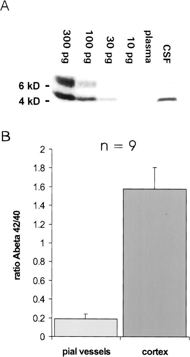
Levels of Aβ in CSF and plasma, and the ratio of insoluble Aβ42:Aβ40 in pial vessels and neocortex. A: Western blot for Aβ in CSF (5 μl) and plasma (5 μl) from a 4-month-old APP/Ld mouse. Aβ could be detected in CSF (level ≈ 20 pg/μl), but not in plasma. Using a longer exposure time (not shown), it was possible with this method to detect 10 pg of Aβ, but in plasma Aβ remained undetectable (level < 2pg/μl). The band above the 6-kd marker probably represents polymerized Aβ. B: The ratio of Aβ42:Aβ40 is eight times lower in vascular amyloid (0.19 ± 0.04) than in plaque amyloid (1.58 ± 0.23, P = 0.003), showing that Aβ40 is much more abundant relative to Aβ42 in vascular amyloid.